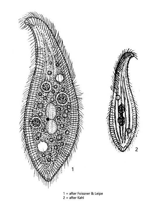
ventrally and dorsally broad seam of extrusomes, not aggregated to warts
right side flat and ciliated
left side naked with 3 – 8 longitudinal ridges
two macronuclear nodules and a spherical micronucleus between them
two contractile vacuoles in anterior and posterior third of cell
extrusomes 6–8 µm long, slightly curved
Siroloxophyllum utriculariae
This ciliate was first described by Penard as Amphileptus utriculariae (1922) and later transferred to Loxophyllum by Kahl (1926) because he saw the naked left side and the fringe of extrusomes running along both the ventral and dorsal margins. Later, the species was then transferred to Spiroloxophyllum by Foissner & Leip (1995), based on the shape of the dorsal brush and a ribbed structure of the ventral and dorsal margins.
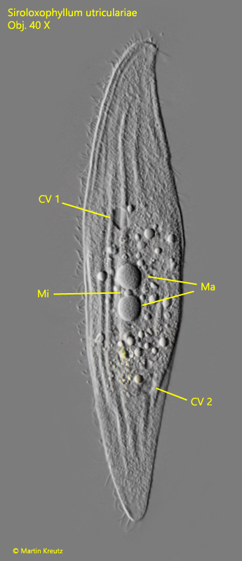
I have found Siroloxophyllum utriculariae only once in August 2003 in the mill pond of Litzelstetten, between floating plants. Unfortunately I took only one photo at that time (s. fig. 1). Nevertheless the main features are visible. The left side shows distinct ridges and the two diagonally vesetted contractile vacuoles in the anterior and posterior third of the body are visible. The macronucleus consists of two parts with a small micronucleus between them. The ciliate is very hyaline.
Siroloxophyllum utriculariae can be distinguished from Loxophyllum meleagrisand Loxophyllum helus because these species have only one contractile vacuole and because the dorsal margin is covered by warty bundles of extrusomes. Siroloxophyllum utriculariae is distinguished from Litonotus species by the extrusomal fringe on the ventral and dorsal margins (only ventral and terminal in Litonotus) and the number of contractile vacuoles (only one terminal CV in Litonotus).
Fig. 1: Siroloxophyllum utriculariae. L = 212 µm. A freely swimming specimen from left. Note the longitudinal ridges of the left side. CV 1, CV 2 = contracile vacuoles; Ma = macronuclear nodules; Mi = micronucleus. Obj. 40 X.