body medusoid with a primary spine and a secondary spine
primary spine swollen in the middle and with an additional, affiliated spine at distal end
secondary spine thin, slightly curved
length 50–100 µm
one spherical macronucleus with a small adjacent micronucleus
on left side of the dome one row of cilia
acculumation of refractive granules in apical dome (sometimes absent)
without somatic cilia except a field of cilia at the base of the primary spine
adoral zone runs in a furrow encircling the body spirally
mouth opening in mid-body, cytopharynx directed anteriorly
different species of symbiotic bacteria in the cytoplasm
one contractile vacuole located at the base of the primary spine
Sulfonecta uniserialis
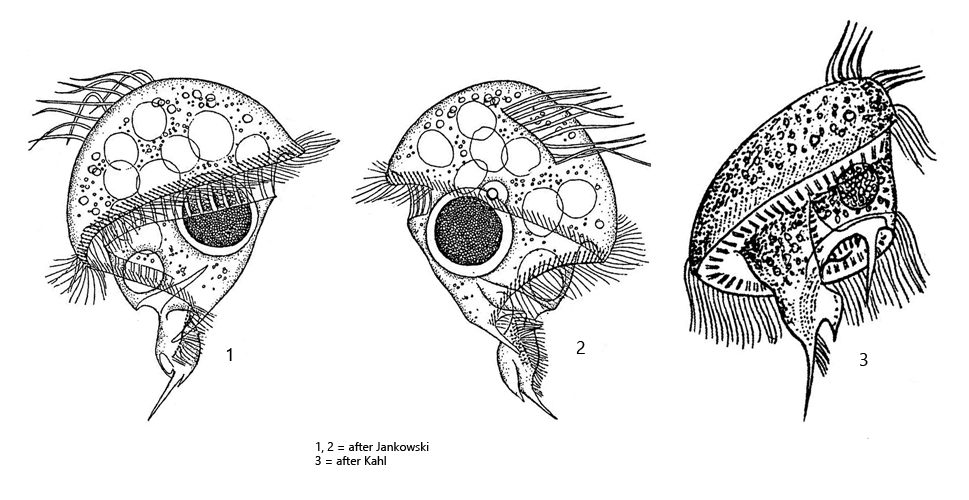
Sulfonecta uniserialis was first described by Levander as Caenomorpha uniserialis. This name was also adopted by Kahl. Later Caenomorpha uniserialis was transferred to the genus Sulfonecta by Jankowski. It is the only species in the genus Sulfonecta. We will see if this remains to be the case.
In my localities Purren pond, Ulmisried and Simmelried I find Sulfonecta uniserialis frequently and regularly in the mud zone. The species has two caudal spines, but they are comparatively short in relation to the body length. The broad and flat apical dome makes Sulfonecta uniserialis look almost somewhat lenticular at low magnifications. The centrally located primary spine is conspicuously swollen in the middle or last third. I have never found specimens without this swelling. In addition, this primary spine still has an accessory spine, which was always short and pointed in my population (s. fig. 2 b). The secondary spine is much shorter and somewhat displaced towards the outer margin (s. figs. 2 c and 3 b). In my population it was always slightly curved towards the central long axis. As in Caenomorpha medusula and Caenomorpha sapropelica, the cytopharynx is situated near the contractile vacuole and oriented toward the anterior end (s. fig. 1 c). Unlike these two species, Sulfonecta uniserialis has only one row of cilia on the apical dome instead of two. This row of cilia is located on the left side and is difficult to recognize. According to Kahl, this row of cilia is supposed to be soft cirri, which disintegrate due to coverslip pressure. The macronucleus is large and spherical, while the micronucleus is exceptionally small and attached to the macronucleus (s. fig. 3 b). I was able to detect symbiotic bacteria in the cytoplasm of all investigated specimens (s. fig. 3 a-b). Although I did not examine these more closely, several different forms (species) of bacteria were clearly visible.
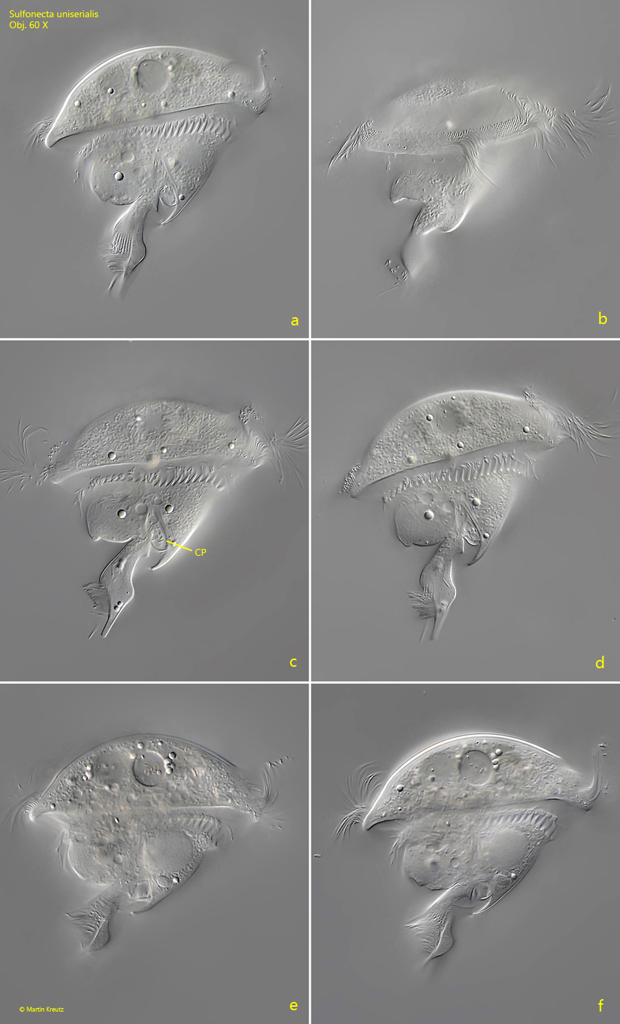
Fig. 1 a-f:Sulfonecta uniserialis. L = 86 µm. Different focal planes of a freely swimming specimen from right. Note the cytopharynx (CP) directed to the anterior end. Obj. 60 X.
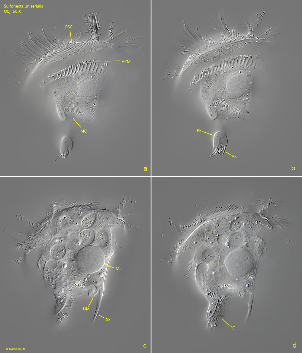
Fig. 2 a-d:Sulfonecta uniserialis. L = 82 µm. Different focal planes of a slightly squashed specimen from left. AS = accessory spine of the primary spine, AZM = adoral zone of membranelles, FC = field of cilia at the base of the primary spine, Ma = macronucleus, MO = mouth opening, PS = primary spine, PSC = perizonal stripe of cilia, SS = secondary spine, UM = undulating membrane. Obj. 60 X.
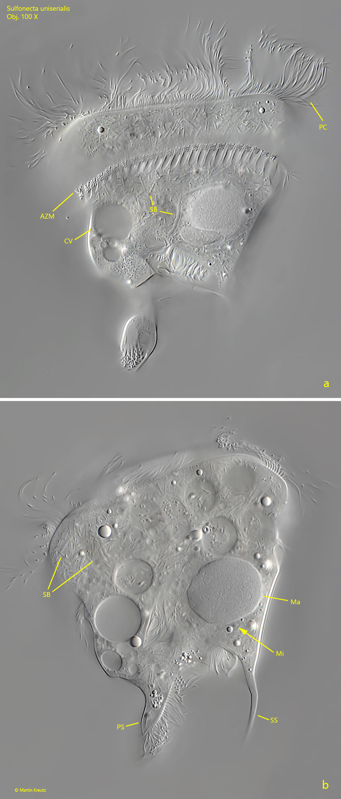
Fig. 3 a-b:Sulfonecta uniserialis. Focal planes on the adoral zone (AZM, a) and macronucleus (Ma, b) of a squashed specimen. Note the small, spherical micronucleus (Mi) adjacent to the globular macronucleus (Ma). In the cytoplasm different species of symbiotic bacteria (SB) are visible. CV = contractile vacuole, PC = perizonal cilia, PS = primary spine, SS = secondary spine. Obj. 100 X.