head and neck contractile pellicle ribbed with long cilia
vividly movement
phyryngeal extrusomes delicate rods
macronucleus oval or kidney-shaped, with one micronucleus
contractile vacuole terminal
Lacrymaria sapropelica
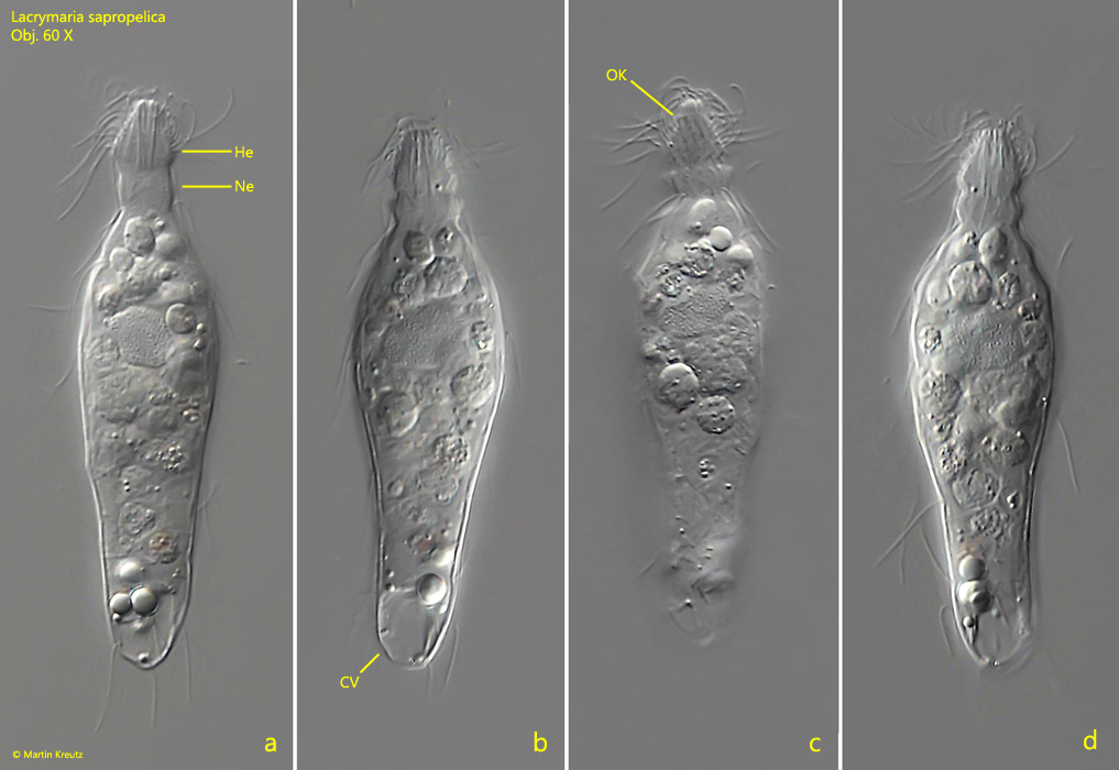
I find Lacrymaria sapropelica in the anaerobic sludge of the Simmelried rarely, but regularly and at longer intervals. This species can be recognized by the fact that in the extended specimen the head is wider than the neck (s. figs. 1a and 2a). The head and neck show striping due to obliquely running kineties, which are difficult to see in the fast-moving specimens. Only in the flash photos (s. fig. 1c) or in the squashed specimen does this become clearly visible.
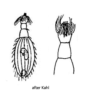
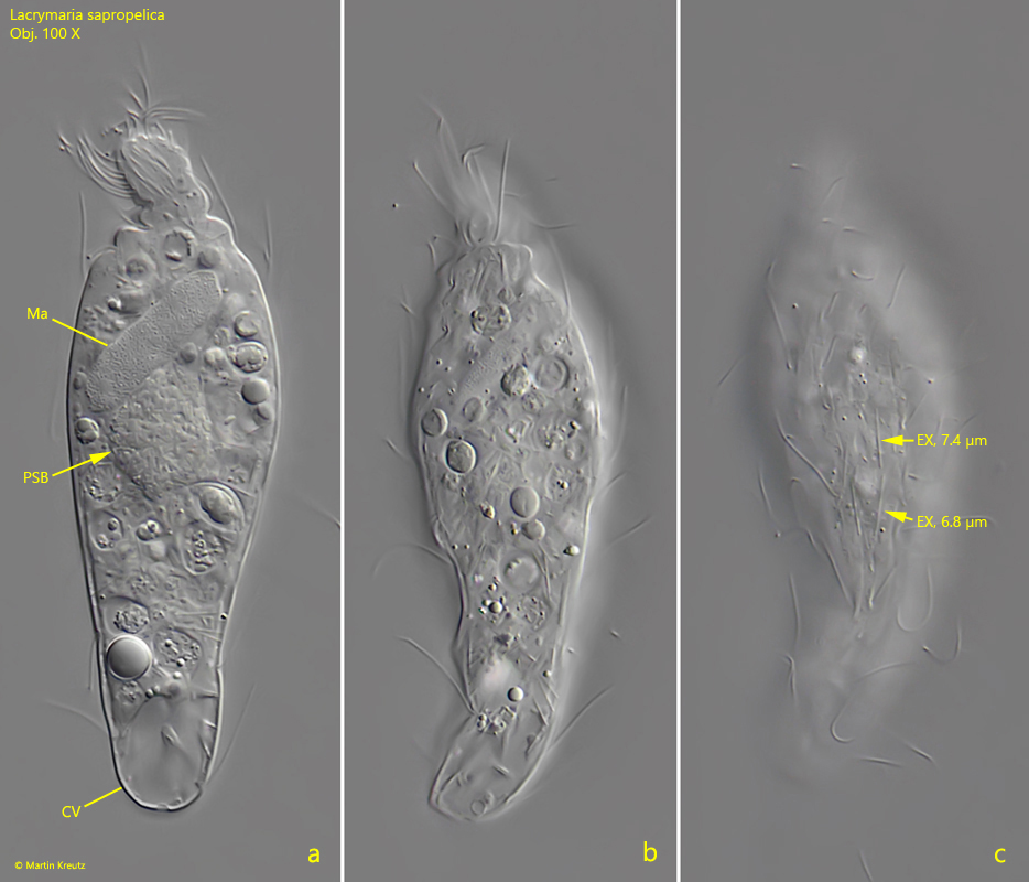
The extrusomes are thin rods, which in my specimens have a length of 6.8–7.4 (s. fig. 3 c) µm. They were also found distributed in the cytoplasm. Although Kahl drew this species with an oval body (s. drawing above), all specimens I found had a bottle shape or amphora shape. The macronucleus in my specimens was always ellipsoid or elongate ellipsoid and was always located in the anterior half of the body (s. figs. 2 b and 3 a). I could not detect the micronucleus.
In 1991, Lacrymaria sapropelica was investigated by Finlay et al. They were able to detect a pouch filled with symbiotic, methanogenic bacteria, which lies directly adjacent to the macronucleus. I was also able to clearly see this pouch with bacteria (s. fig. 3a), which is why I consider the identification of Lacrymaria sapropelica to be very certain.
Fig. 1 a-d:Lacrymaria sapropelica. L = 85 µm. A freely swimming specimen. In the elongated form (fig. 1a) the head (He) is broader than the neck (Ne). Head and neck are show oblique kineties (OK). Obj. 60 X.
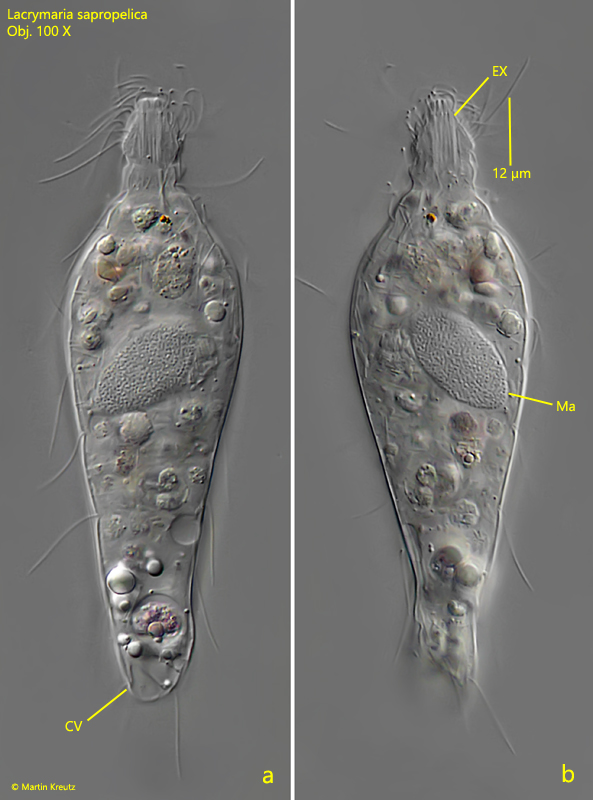
Fig. 2 a-b:Lacrymaria sapropelica. L = 95 µm. A second freely swimming specimen. CV = contractile vacule, EX = extrusomes, Ma = macronucleus. Obj. 100 X.
Fig. 3 a-c:Lacrymaria sapropelica. L = 88 µm. Three focal planes of a third, slightly squashed specimen. The extrusomes (EX) scattered in the cytoplasm have a length of 6.8–7.4 µm. Adjacent to the macronucleus a pouch filled with symbiotic bacteria (PSB) is visible. CV = contractile vacuole, Ma = macronucleus. Obj. 100 X.