body slenderly oval, broadly oval or pyriform, depending on nutritional state
length 60–80 µm
cell surface cross-rippled between the rows of cilia
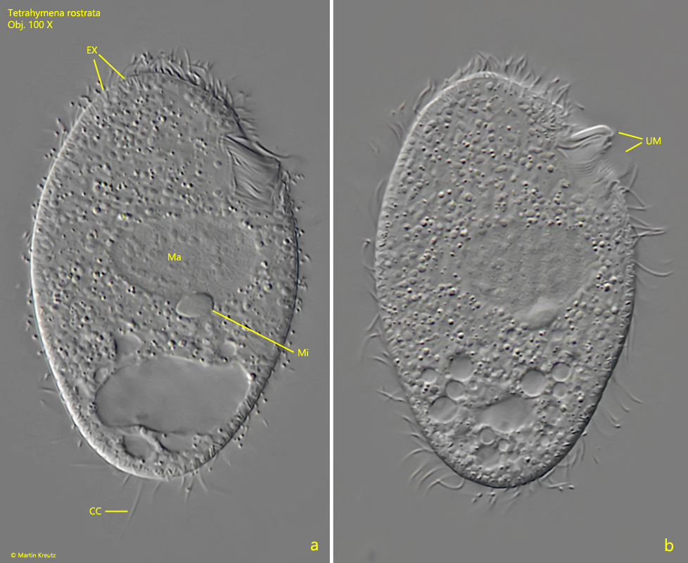
oral apparatus in anterior fifth, with undulating membrane at right side
cilia rows preorally roof-shaped
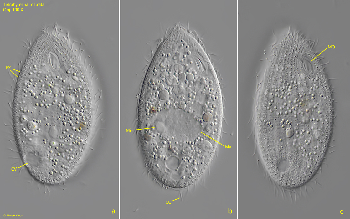
macronucleus oval in mid-body with adjacent spherical micronucleus
contractile vacuole at posterior sixth, located ventrally on the right side
fringe of inconspicuous extrusomes, about 2 µm long
a short, bristle-like caudal cilium
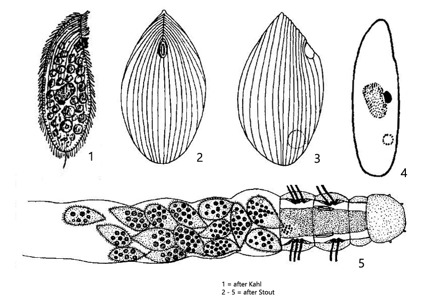
Tetrahymena rostrata
Tetrahymena rostrata was first described by Kahl as Paraglaucoma rostrata and later assigned to Tetrahymena by Corliss. The identification of Tetrahymena rostrata in field samples is difficult given the life cycle of this ciliate. It is facultatively parasitic in slugs (e.g. Deroceras reticulatum) or grazes on dead rotifers and insect larvae (s. fig. 4 a-b). During life cycle, Tetrahymena rostrata passes through different developmental stages and assumes different body shapes (slender-oval and transparent to pear-shaped and opaque) depending on its nutritional state. In addition to this variability, Tetrahymena rostrata is also easily confused with Glaucoma maupasi. However, the latter species has no caudal cilium and the contractile vacuole on the left side.
Tetrahymena rostrata is best identified by the transverse striation of the pellicle (s. fig. 1e), the caudal cilium and the mouth opening in the anterior fifth with an undulating membrane on the right side (s. fig. 3b).
Fig. 1 a-e:Tetrahymena rostrata. L = 74 µm. Different focal planes of a freely swimming specimen. Note the cross-rippled pellicle (fig. 1e). CC = caudal cilium. Obj. 60 X.
Fig. 2 a-c:Tetrahymena rostrata. L = 54 µm. Different focal planes of a slightly squashed specimen. CC = caudal cilium, CV = contractile vacuole, EX = fringe of extrusomes, Ma = macronucleus, Mi = micronucleus, MO = mouth opening. Obj. 100 X.
Fig. 3 a-b:Tetrahymena rostrata. Two focal planes of a strongly squashed specimen from the right side. CC = caudal cilium, EX = fringe of extrusomes, Ma = macronucleus, Mi = micronucleus, UM = undulating membrane at right side of mouth opening. Obj. 100 X.
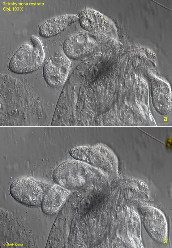
Fig. 4 a-b:Tetrahymena rostrata. A cluster of specimens is feeding on an injured mosquito larva. Obj. 100 X.