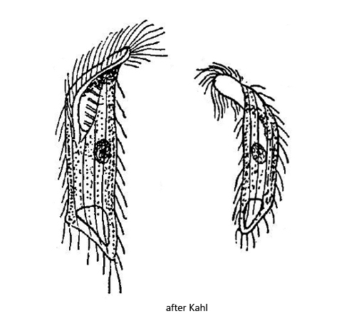
body cylindrical, parallel-sided, ventrally curved
posterior end obliquely truncated
ventral side keeled
length 50–60 µm
adoral zone runs obliquely to the mid-body
cilia loosely arranged, not elongated
macronucleus spherical
contractile vacuole terminal in ventral keel
Metopus ridiculus
Apart from Kahl’s very brief description, I have not found any further information about Metopus ridiculus. He describes the species as “not common” and from his description one can gather that he was not able to examine Metopus ridiculus in detail, since he writes, for example, “(adoral) zone probably up to half”.
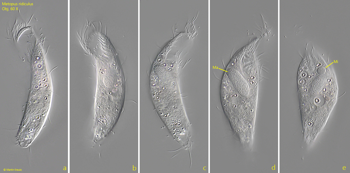
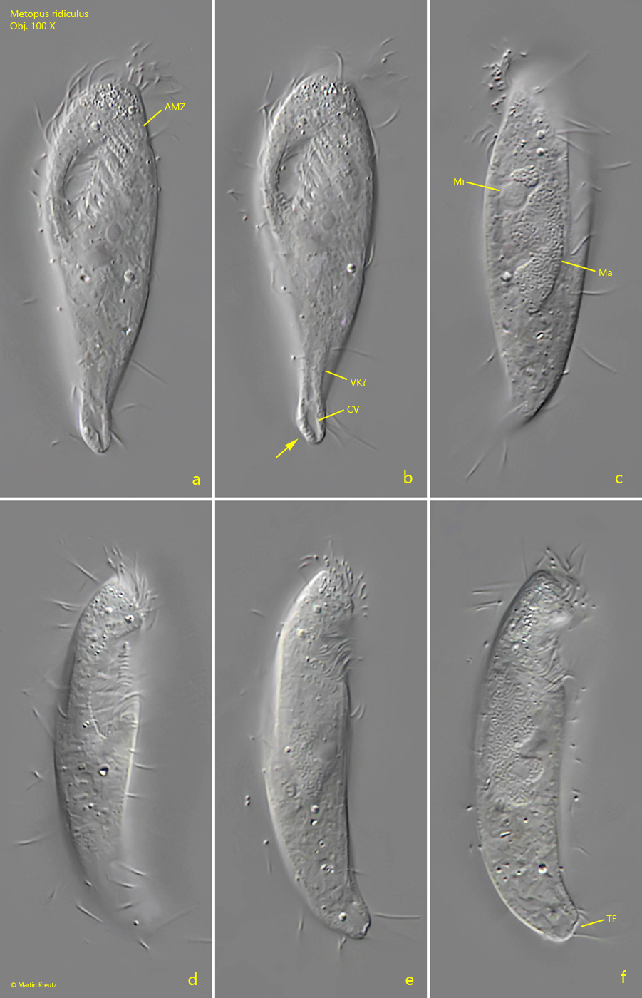
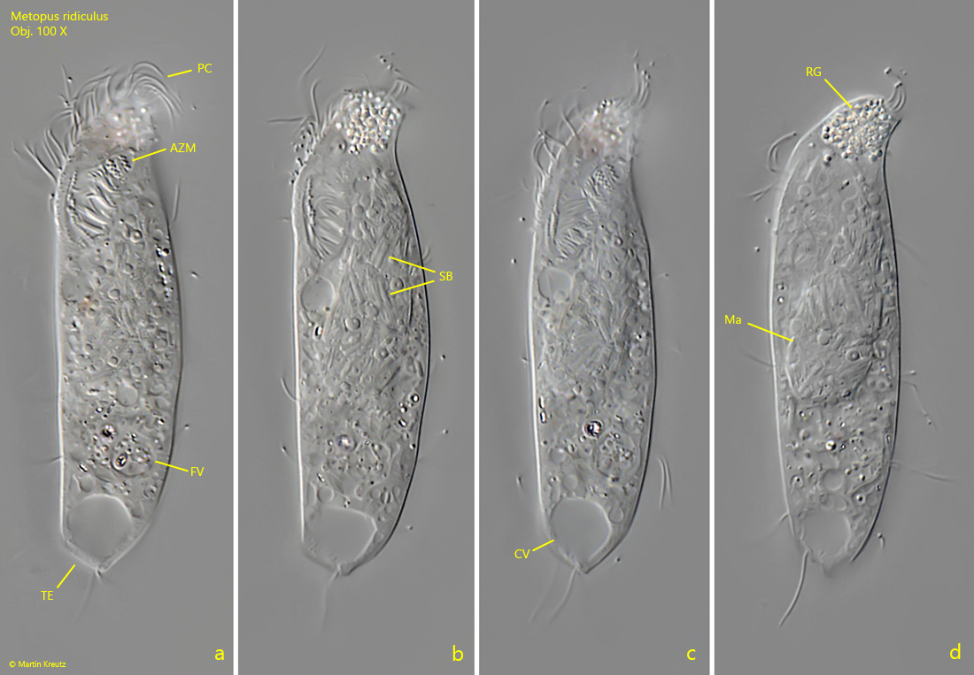
So far I have only been able to find three specimens of Metopus ridiculus. In two of these specimens the body was slightly curved. Only in one specimen was the body parallelsided, as described by Kahl. The truncated posterior end could be clearly seen in 2 specimens (s. figs. 2 a-f and 3 a-d). In one specimen it was only suggested. In all specimens the adoral zone extended to one fifth or one quarter of the body length. In no case did it reach the middle of the body.
The ventral keel mentioned by Kahl I could only recognize in rudimentary form. The body length of my specimens was between 50–75 µm and thus within the range given by Kahl. The macronucleus in my specimens was oval or elliptical with a large, attached micronucleus. In one specimen the macronucleus was permeated by symbiotic bacteria (s. fig. 3 a–d). In the other two specimens this was not the case; however, numerous symbiotic bacteria were scattered in the cytoplasm of these specimens. The contractile vacuole is terminal. I could not, however, determine precisely whether it is located in the ventral keel, as Kahl describes.
Fig. 1 a-e:Metopus ridiculus. L = 75 µm. A a freely swimming specimen found in October 2025 in the Purren pond. Obj. 60 X.
Fig. 2 a-f:Metopus ridiculus. L = 50 µm. A a freely swimming specimen from ventral (a, b), from left (c) and from right (d – f). Note the truncated posterior end from ventral (arrow, b) and from left (TE). AMZ = adoral zone of membranelles, CV = contractile vacuole, Ma= macronucleus, Mi = micronucleus, VK? = likely the ventral keel. Obj. 100 X.
Fig. 3 a-d:Metopus ridiculus. L = 62 µm. Different focal planes of a slightly squashed specimen from ventral. AMZ = adoral zone of membranelles, CV = contractile vacuole, Ma= macronucleus, PC = perizonal cilia, RG = aggregation of refractive granules in the apical dome, SB = symbiotic bacteria. Obj. 100 X.