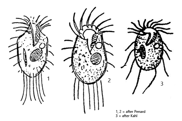
oral apparatus at anterior end with an inner zone of 5–10 membranelles
in equatorial zone bifurcated jumping bristles
several long trailing cilia at posterior end
cytoplasm sometimes with few symbiotic algae
macronucleus ovoid with adjacent micronucleus, located centrally
one contractile vacuole left to the mouth opening
lives in the gelatinous mass of Chaetophora
Halteria oblonga
Both Penard (1922) and Kahl (1932) found Halteria oblonga exclusively in the gelatinous mass of the green alga Chaetophora. I can confirm this finding (s. fig. 1). If freely swimming specimens are found, they were released from the jelly by the sample preparation. So far I have only been able to find Halteria oblonga in the Simmelried, where the species is rarely found.
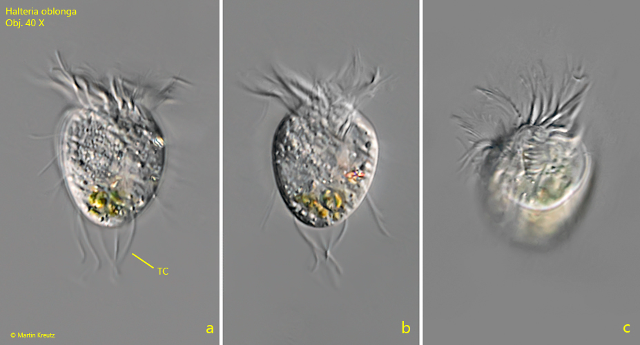
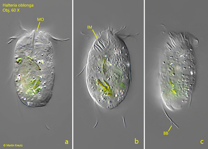
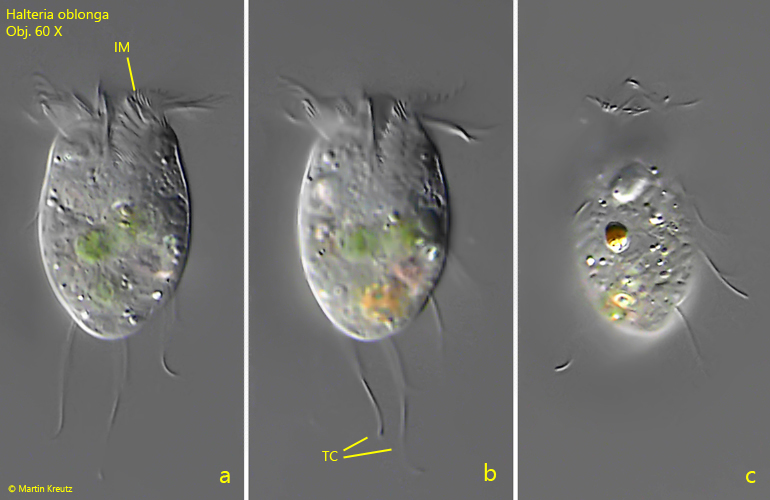
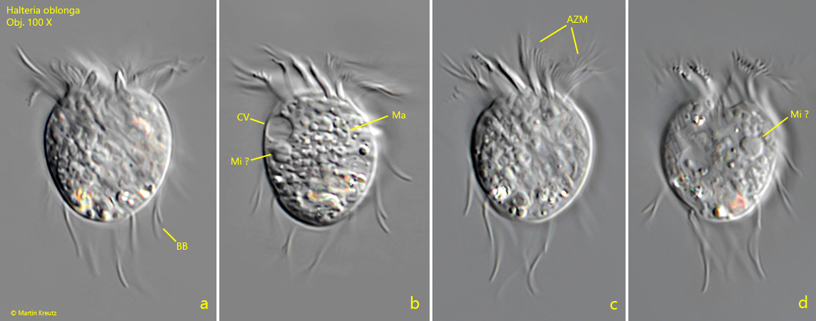
Halteria oblonga has spring bristles, which are bifurcated at the distal end (s. figs. 3 c and 5 a). However, these appear to be soft and flexible, as they are kept attached to the body during swimming. About 4–6 trailing cilia, which often have a characteristic S-shaped form, protrude beyond the posterior (s. figs. 1 a and 4 b). The outer adoral zone of membranelles (s. fig. 5 c) is not as strongly developed as in the species Halteria grandinella. The inner zone of membranelles (s. figs. 3 b and 4 a), which runs to the mouth opening, does not reach a fourth of the body length and runs slightly to the right. The body shape seems to be quite variable from broadly oval (s. fig. 5 a-d) to almost ellipsoid (s. fig. 4 a-c). I have not yet found specimens with symbiotic algae in the cytoplasm, as described by Kahl.
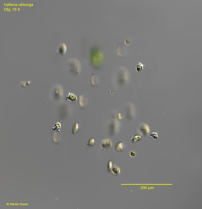
Fig. 1:Halteria oblonga. Several specimens in a piece of gelatinous mass of the algae Chaetophora. Obj. 10 X.
Fig. 2 a-c:Halteria oblonga. L = 43 µm. Lateral (a, b) and apical view of a freely swimming specimen. Note the tuft of trailing cilia (TC). Obj. 40 X.
Fig. 3 a-c:Halteria oblonga. L = 42 µm. An almost ellipsoidal specimen. Note the bifurcated jumping bristles (BB). IM = inner membrelles of the adoral zone, Mo = mouth opening. Obj. 60 X.
Fig. 4 a-c:Halteria oblonga. L = 42 µm. An oval specimen with long, S-shaped trailing cilia (TC). IM = inner membrelles of the adoral zone. Obj. 60 X.
Fig. 5 a-d:Halteria oblonga. L = 42 µm. An broadly oval specimen. AZM = adoral zone of membranelles, BB = bifurcated jumping bristle, CV = contractile vacuole, Ma = macronucleus, Mi ? = probably the micronucleus. Obj. 100 X.