two reddish blisters between intestinum and bladder
stomach and intestine colored golden brown or orange
foot very short and stout
toes extremely long and unequal (150–400 µm long)
Monommata actices
I only find Monommata actices in the Simmelried where the species is quite common. I mainly find specimens between floating plant masses. In the sample vessel they gather at the surface.
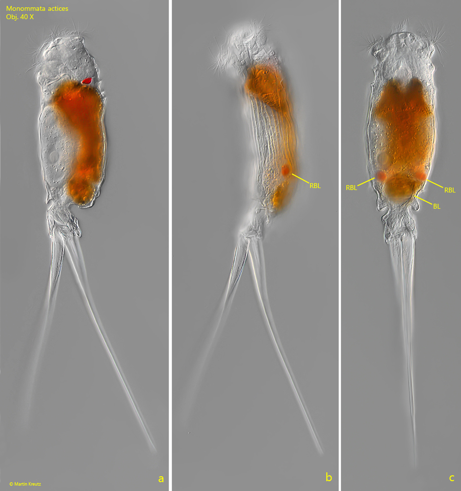
Monommata actices has an eyespot, which is located at the posterior end of the ganglion (s. fig. 4). The cuticle is clearly longitudinally striated (s. figs. 1 b and 2 b) and between the intestine and the bladder is a pair of reddish blisters is located whose function is unknown (s. figs. 1 b, 1 c, 2 a and 5). At higher magnification it can be seen that these reddish blisters contain a central, reddish oil body (s. fig. 5). They could be glands that produce a reddish secretion. The stomach and intestines are always golden brown or orange in color due to ingested Synura colonies. This was the case with all the specimens I examined. The two extremely long toes are of different lengths and contain striated muscle cells. With the help of these toes, Monommata actices can perform jumps quickly.
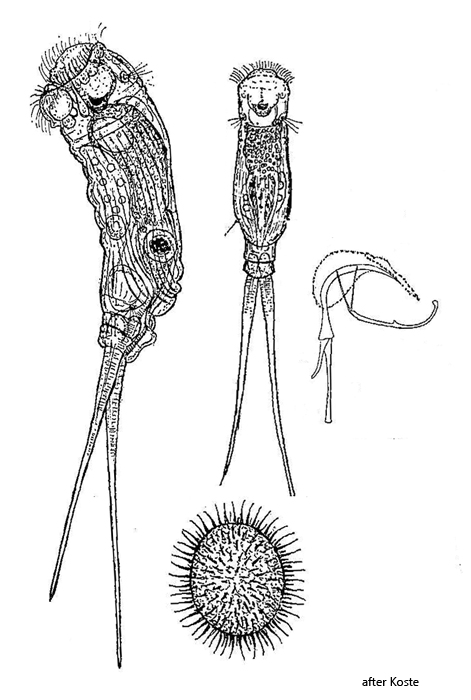
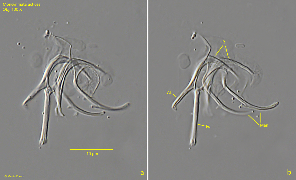
The unambiguous identification of species within the genus Monommata is only possible with the trophi. To prepare this, isolated specimens must be treated with an approx. 1% SDS solution (SDS = sodium-dodecyl-sulfate). This macerates and exposes the trophi (s. fig. 7 a-b). On the trophi of Monommata actices the the alula can be recognized, which emerges from the fulcrum. This is typical for the species (compare to drawing above).
Fig. 1 a-c:Monommata actices. L = 455 µm (with toes). A freely swimming specimen from left (a, b) and from dorsal. Note the longitudinal striation of the cuticle (b) and the pair of reddish colored blisters (RBL) located between intestine and bladder (BL). Obj. 40 X.
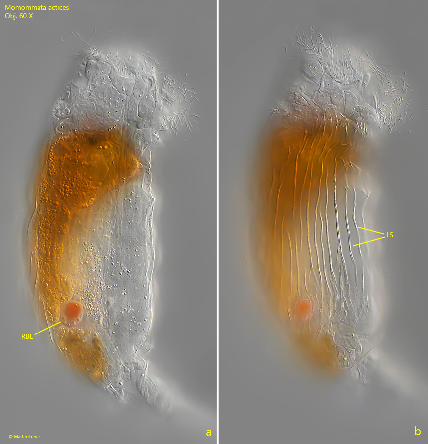
Fig. 2 a-b:Monommata actices. Two focal planes of a slightly squashed specimen. Note the reddish blister (RBL) and the longitudinal striation (LS) of the cuticle. Obj. 60 X.
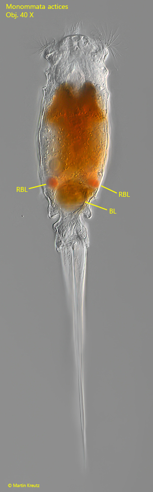
Fig. 3:Monommata actices. A specimen from dorsal with two reddish blisters (RBL). BL = bladder. Obj. 40 X.
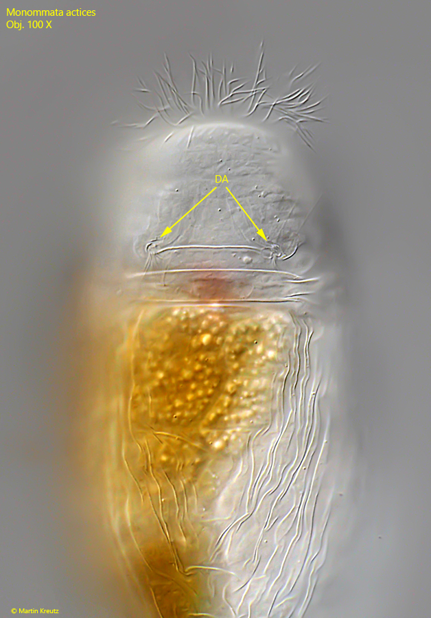
Fig. 4:Monommata actices. Dorsal view of a slightly squashed specimen with focal plane on the both dorsal antennae (DA). Obj. 100 X.
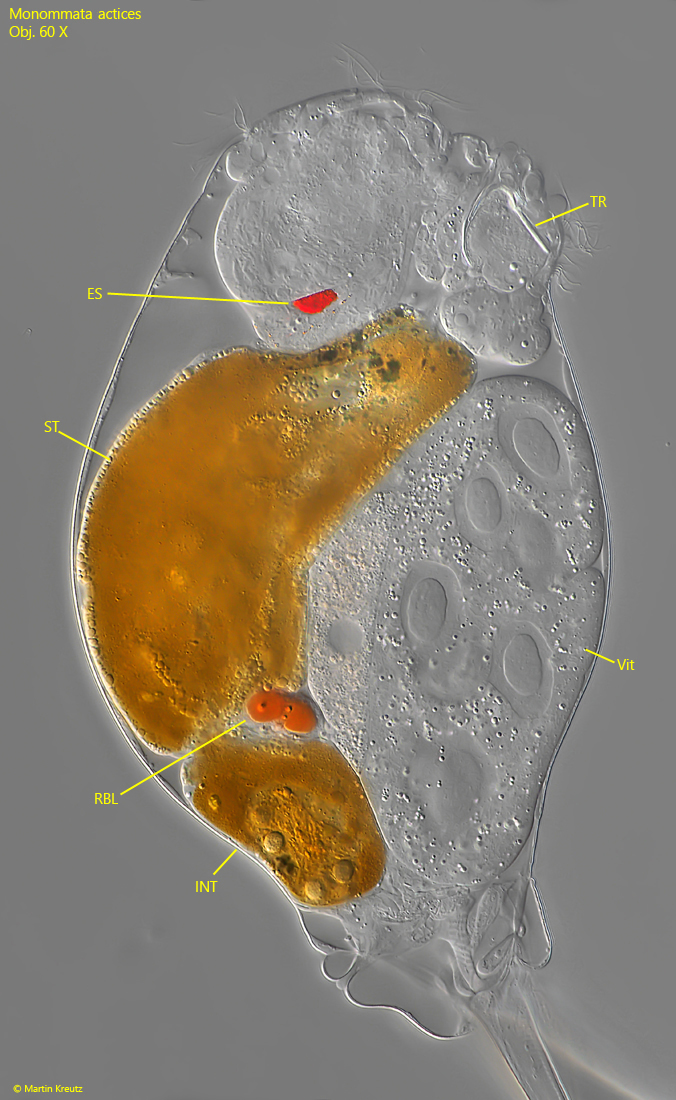
Fig. 5:Monommata actices. A strongly squashed specimen from right. ES = eyespot, INT = intestine, RBL = reddish blister, ST = stomach, TR = trophi, Vit = vitellarium. Obj. 60 X
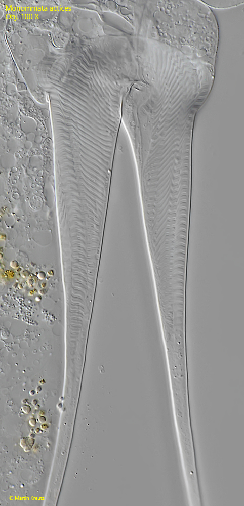
Fig. 6:Monommata actices. The striated muscles in the toes. Obj. 100 X
Fig. 7 a-b:Monommata actices. Two focal planes of the macerated trophi in a strongly squashed specimen. AL = alula, FU = fulcrum, Man = manubria, R = rami. Obj. 100 X.
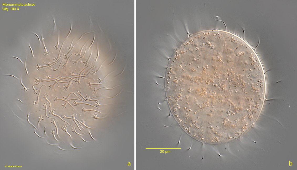
Fig. 8 a-b:Monommata actices. Two focal planes of an amictic egg. Obj. 100 X.