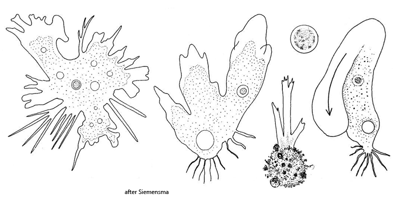
polypodial form fan-shaped, with numerous pseudopodia
monopodial form elongated ovoid, finger-shaped
pseudopodia with hyaline cap
length polypodial form 35–60 µm
length monopodial form 42–100 µm
nucleus 6.5–8.0 µm, with a central, spherical nucleus
one contractile vacuole
cytoplasm clear, few food vacuoles, crystals absent
uroid filmentous
Rhizamoeba timidum
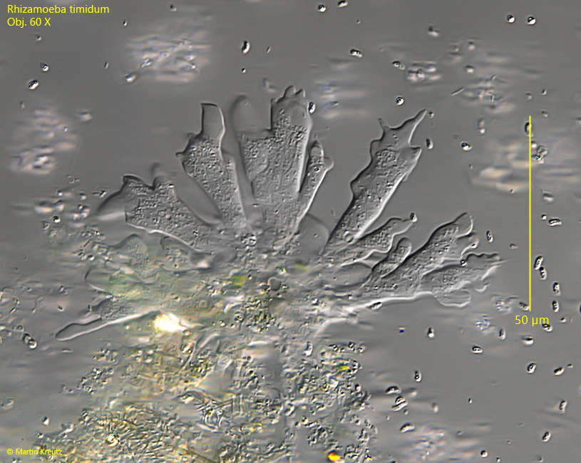
I have only been able to find Rhizamoeba timidum in the Simmelried so far, but only until 2014, after which I have not found any more specimens. The specimens shown below are from a population from March 2010.
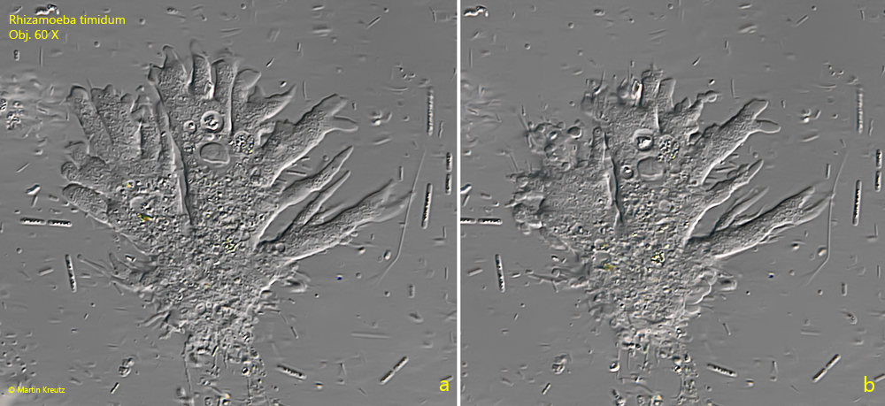
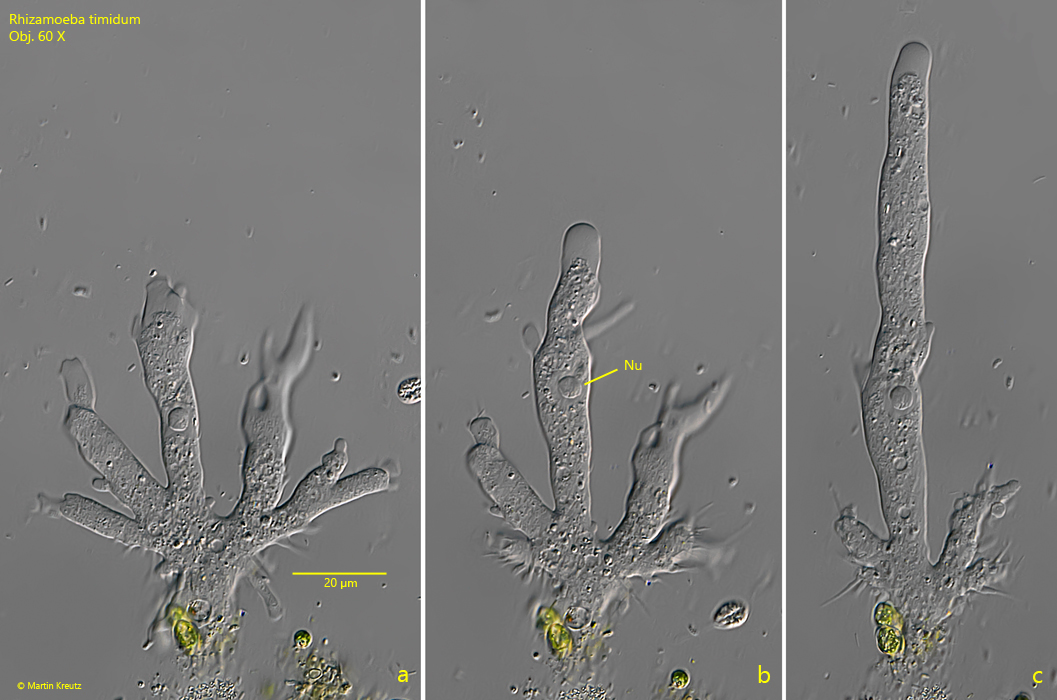
The polypodial form of Rhizamoeba timidum is fan-shaped and strongly flattened. The specimens are often covered with detritus at the posterior end of the body and are stuck in detritus. The pseudopodia often show finger-shaped protrusions that are hyaline. This gives Rhizamoeba timidium a typical appearance. However, an exact classification of the species is only possible by the shape and size of the nucleus. In Rhizamoba timidium, the nucleus is about 6-8 µm in size and has a homogeneous, spherical nucleolus (s. fig. 3 b).
Rhizamoeba timidium appears to be sensitive to light or heat radiation. As soon as an outstretched specimen is moved into the illuminated part of the slide, contraction begins very quickly. Although my microscope is equipped with a heat protection filter and I tried to work with minimal light intensity, this effect always occurred. I was able to document the light sensitivity of Rhizamoeba timidum well in a video (sorry, it’s in German):
Fig. 1:Rhizamoeba timidum. L = 58 µm. A specimen in the polypodial form which is partly covered with detritus. Obj. 60 X.
Fig. 2 a-b:Rhizamoeba timidum. L = 65 µm. A second specimen in the polypodial form. Obj. 60 X.
Fig. 3 a-c:Rhizamoeba timidum. A specimen during the transition from the polypodial form (a) into the monopodial form (c). The monopodial form has a length of 158 µm. The diameter of the nucleus is 8.3 µm. Obj. 60 X.