I found Distigma sennii in August 2010 in the Simmelried. After that I have no further records. But perhaps I have often overlooked the species because the second, short flagellum, which characterizes the genus Distigma, can only be seen at high magnification.
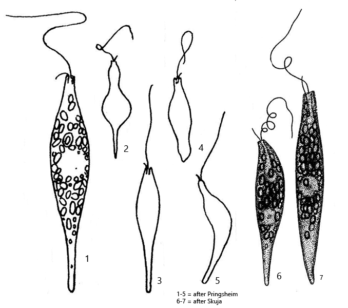
The classification as Distigma sennii is based on the truncated anterior end, the length of the cells of about 50 µm and the conical body shape with a tapered posterior end. It can therefore not be Distigma proteus. This species is about twice as long and has a pronounced striation of the pellicle. Distigma globiferum is smaller and the mid-body is clearly enlarged due to a very large nucleus. A further alternative would be Distigma elegans. This species is about the same length and also has a truncated anterior end, but is described to have a very strong euglenoid movement.
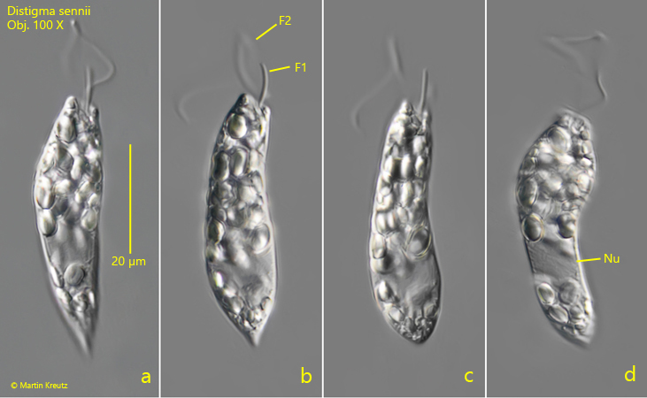
Fig. 1 a-d:Distigma sennii. L = 47 µm. Different phases of the euglenoid movement of a freely swimming specimen. Note the two flagella of different length (F1, F2) and the truncated anterior end. Nu = nucleus. Obj. 100 X.
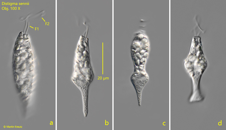
Fig. 2 a-d:Distigma sennii. L = 54 µm. A second specimen during euglenoid movement. Obj. 100 X.