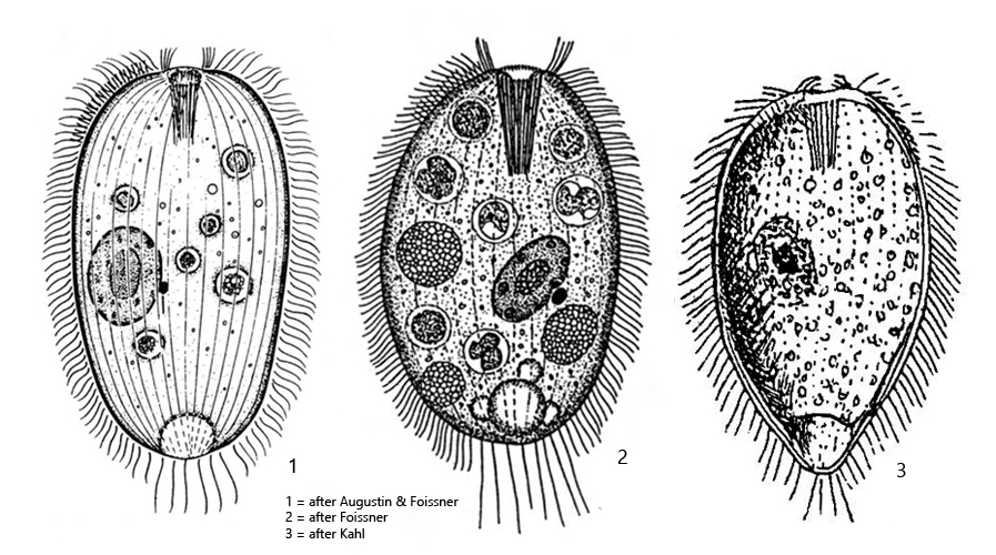
mouth opening apical, oral basket with 25–35 trichites
adoral brush (3 rows)
35–64 longitudinal rows of cilia
macronucleus ellipsoid, one adjacent micronucleus
extrusomes 2–3 µm long, spindel-shaped, sometimes absent
contractile vacuole terminal with several excretion pores
several caudal cilia
Holophrya discolor
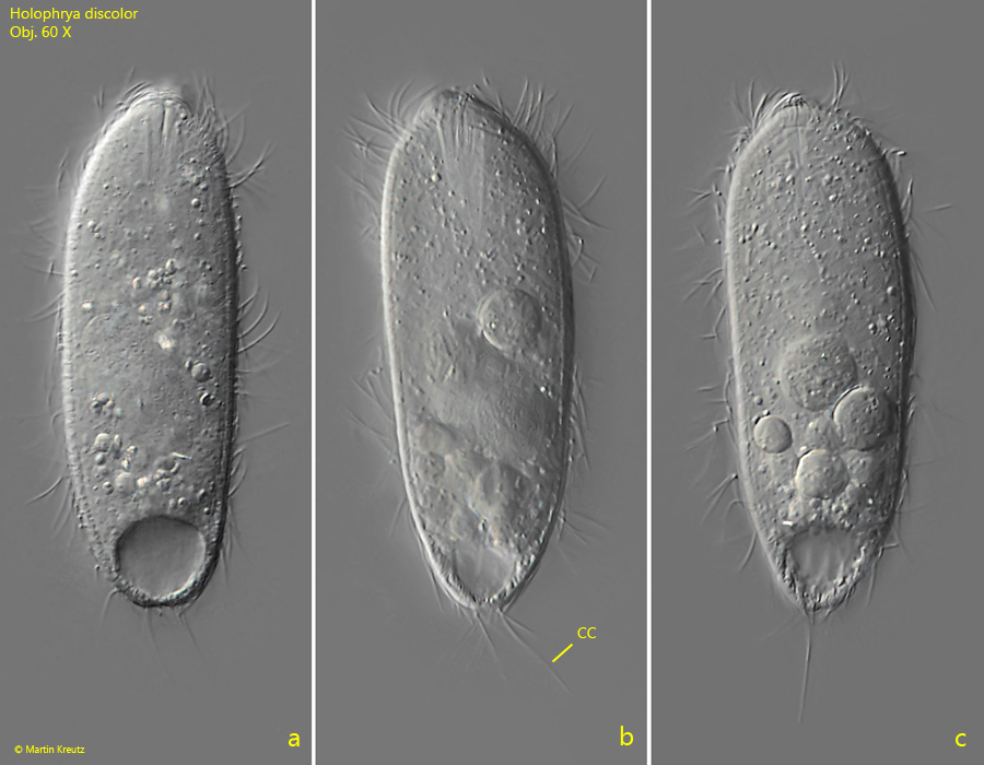
Holophrya discolor is a very common ciliate that occurs in practically all of the sampling sites. The species has a complex life cycle (theront-trophont-protomont-tomont-tomit). The images below show the theront phase, with little ingested food. In the trophont and protomont phases, the specimens are completely filled with food vacuoles, rounded and opaque.
Important characteristics for the classification are the number of longitudinal rows of cilia, the size and the absence of symbiotic algae. The cell surface is covered by 35–60 longitudinal rows of cilia, which are located in furrows. The total number can only be seen in anteriorly or posteriorly view. In lateral view, about 20 longitudinal rows can be recognized in squashed specimens (s. figs. 6 and 7). The similar species Holophrya teres is larger (usually 150–250 µm) and has 80-110 longitudinal rows, i.e. about twice as many as Holophrya discolor. If the characteristics are similar to those of Holophrya discolor, but the cytoplasm contains symbiotic algae, then it is Holophrya ovum.
The adoral brush of Holophrya discolor is dexiotropic, i.e. it runs to the right towards the mouth opening (s. figs. 6 and 7). The pellicle is covered by a cortical alveolar layer which forms a net-like pattern (s. fig. 5a). However, this pattern is not easily recognizable in all specimens and only in weakly squashed specimens. If the coverslip pressure is too strong, the pattern disappears.
Fig. 1 a-c:Holophrya discolor. L = 78 µm. A freely swimming specimen. CC = caudal cilia. Obj. 60 X.
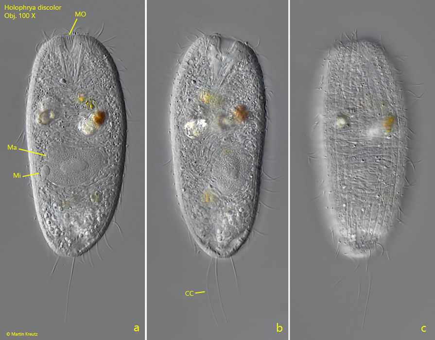
Fig. 2 a-c:Holophrya discolor. L = 81 µm. A second, freely swimming specimen. CC = caudal cilia, Ma = macronucleus, Mi = micronucleus, MO = mouth opening. Obj. 100 X.
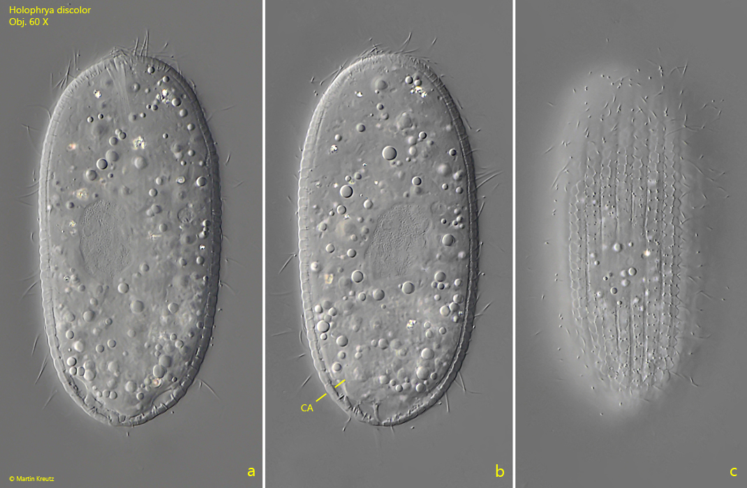
Fig. 3 a-c:Holophrya discolor. L = 103 µm. A third, freely swimming specimen. Note the layer of cortical alveoli (CA). Obj. 60 X.
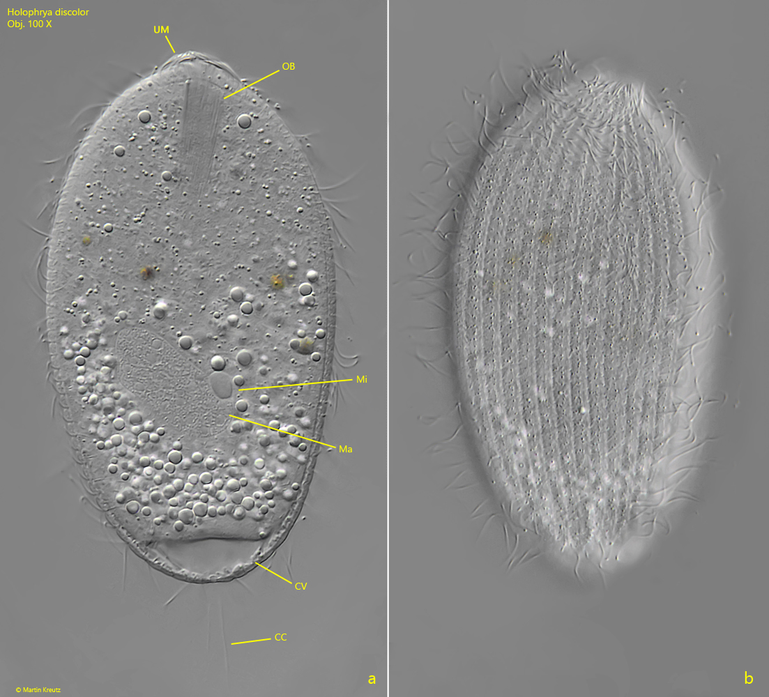
Fig. 4 a-b:Holophrya discolor. Two focal planes of a slightly squashed specimen. Note the cilia of the undulating membrane (UM), which surround the mouth opening in a circle. CC = caudal cilia, CV = contractile vacuole, Ma = macronucleus, Mi =micronucleus, OB = oral basket of trichites. Obj. 100 X.
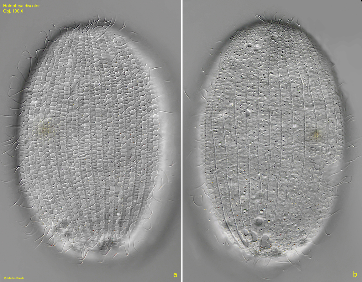
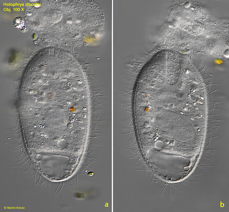
Fig. 5 a-b:Holophrya discolor. The reticulate pattern of the cortical alveoli in a slightly squashed specimen (a). Below the cortical alveoli the pellicle with a longitudinal furrows become visible (b). Obj. 100 X.
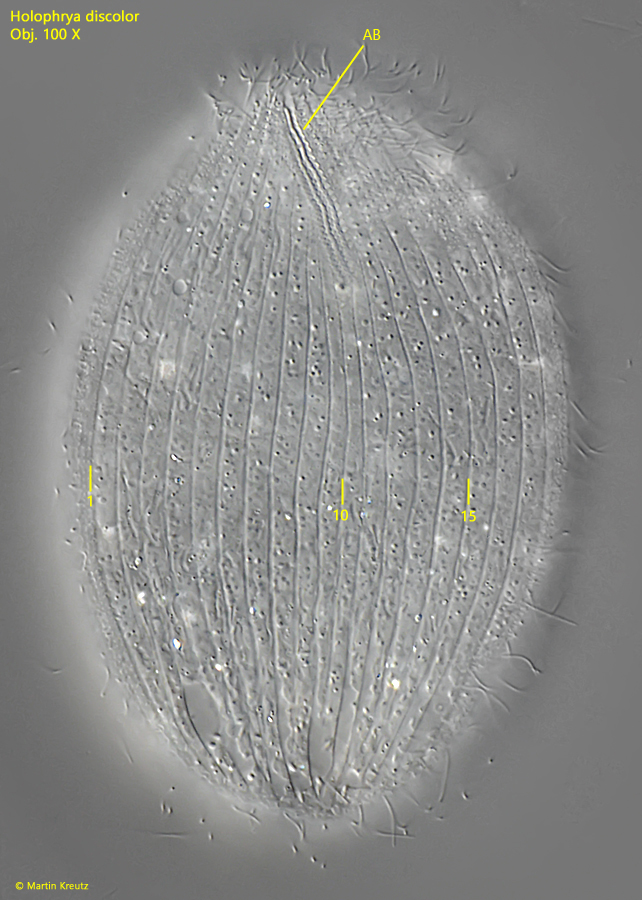
Fig. 6:Holophrya discolor. The adoral brush (AB) in a squashed specimen. Note the oblique course of the adoral brush. It runs to the right towards the mouth opening. This arrangement is referred to as dexiotropic. On this side of the body 18 longitudinal rows of cilia are visible. Obj. 100 X.
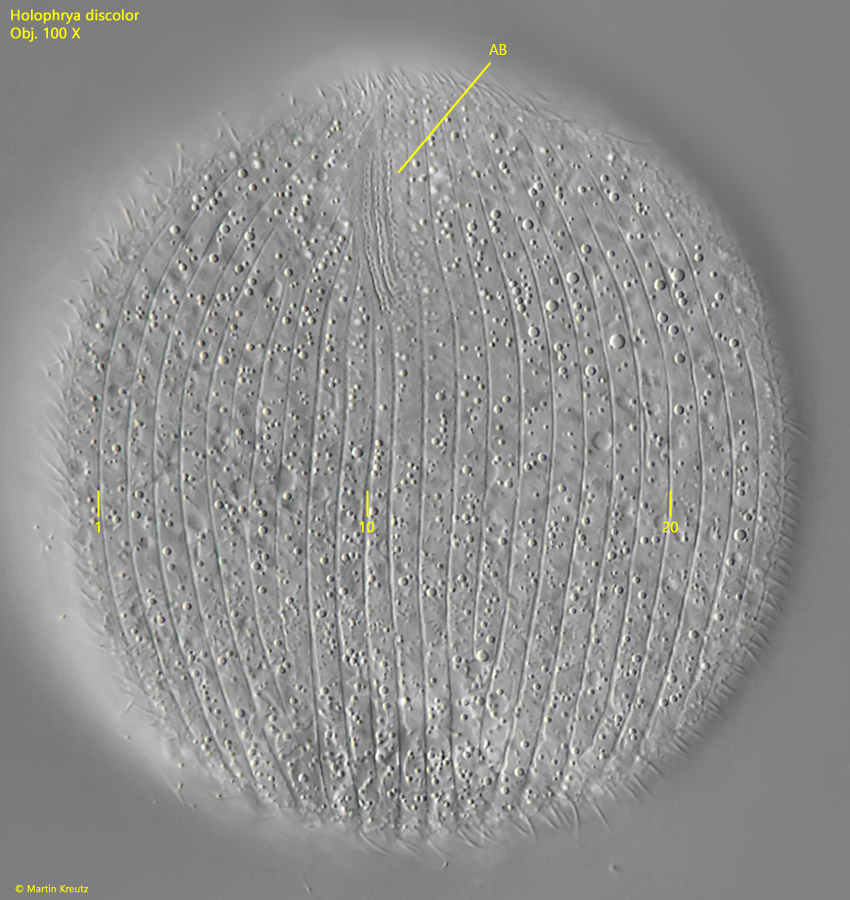
Fig. 7:Holophrya discolor. The adoral brush (AB) in a second, strongly squashed specimen. The number of longitudinal rows of cilia on this side of the body is 23. Obj. 100 X.
Fig. 8 a-b:Holophrya discolor. L = 90 µm. A specimen is ingesting the leaked cytoplasm of a burst ciliates. Obj. 100 X.
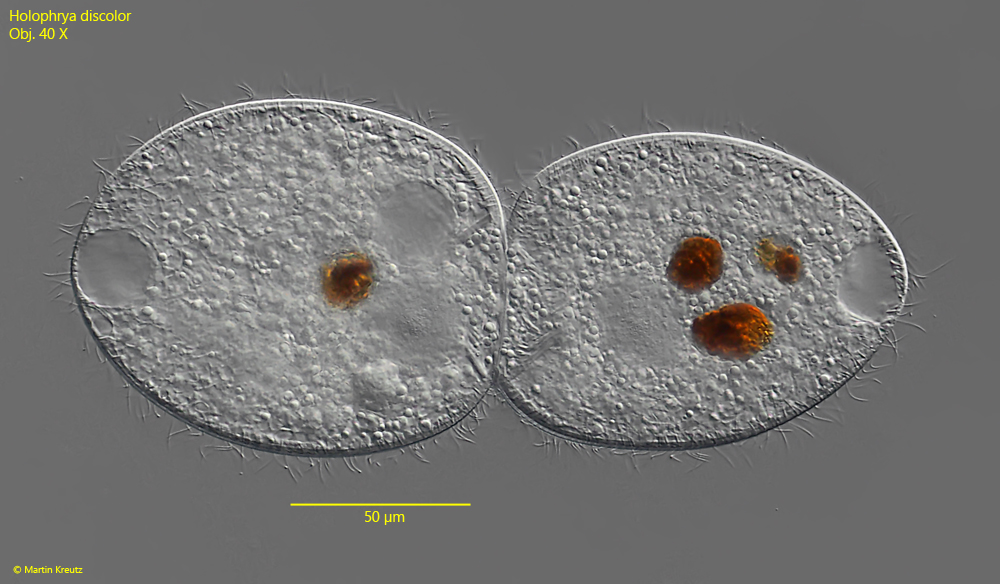
Fig. 9:Holophrya discolor. L = 106–110 µm. Two specimens in conjugation. Obj. 40 X.