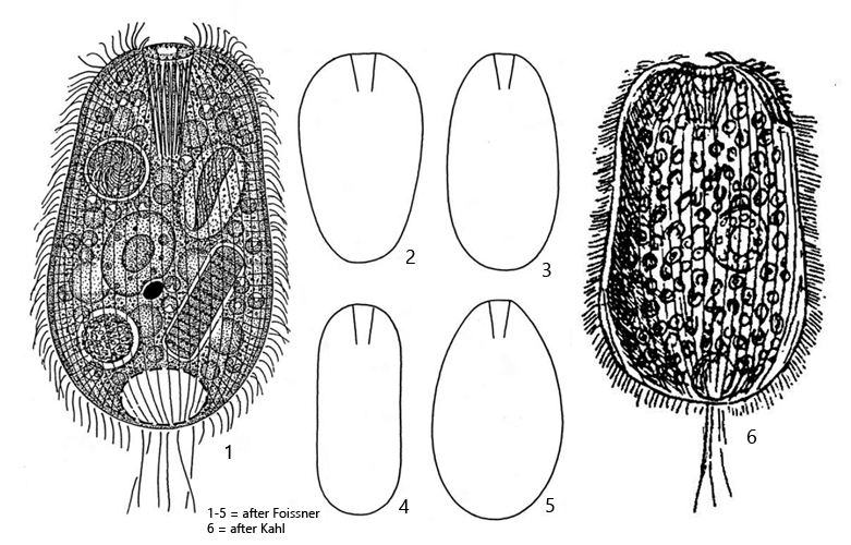
body mostly broadly ellipsoid, slightly constricted in middle, anteriorly obliquely truncated
green due to symbiotic algae
length 100–160 µm
mouth opening apical, oral basket with 24–34 clasp-shaped rods
adoral brush with 3 rows
52–80 longitudinal rows of cilia
macronucleus ellipsoid, one adjacent micronucleus
extrusomes about 8–9 µm long, thin rods, slightly curved
contractile vacuole terminal
several caudal cilia
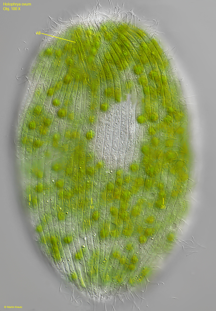
Holophrya ovum
In my sampling sites Holophrya ovum is not very common. In the most cases I found several specimens in old samples with decaying plants. There is a high risk of confusion with specimens of Holophrya discolor, which have phagocytized algae and appears also green. Foissner et al. (1994) explicitly point out this problem. I find such green specimens of Holophrya discolorvery frequently. I can often identify them by their small size (< 100 µm), but estimating the length is difficult, especially at low magnification and roundish specimens. It is therefore possible that I have overlooked other specimens of Holophrya ovum so far.
An important characteristic of Holophrya ovum, apart from the green coloration due to symbiotic algae, is the number of longitudinal rows of cilia and the shape of the extrusomes. The specimens must therefore be examined closely for reliable identification.
The specimens of my population had a length of approximately 90–100 µm and when slightly squashed I was able to count 23 longitudinal rows of cilia on one half of the body (s. fig. 5). For the actual number of longitudinal rows, the lateral rows and those on the opposite side of the body have to be considered. Thus, the number of longitudinal rows of cilia in the specimens of my population was about 50. An important feature is the shape and length of the extrusomes. In Holophrya ovum these are thin, slightly curved rods with a length of 8–9 µm (s. fig. 9). They differ clearly from the extrusomes in Holophrya discolor (2–3 µm, spindle shaped) and Holophrya teres (13–18 µm, thin rods). In my specimen of Holophrya ovum I could clearly detect 9 µm long, slightly curved extrusomes (s. fig. 9). The rods of the basket are clasp-shaped and appear as double-rods (s. fig. 10).
The symbiotic algae of Holophrya ovum are from Chlorella type with a own nucleus. The species was identified as Chlorella faginea by Sud (1969).
Fig. 1 a-d:Holophrya ovum. L = 94 µm. A freely swimming specimen. CC = caudal cilia. Obj. 40 X.
Fig. 2 a-b:Holophrya ovum. L = 100 µm. A second freely swimming specimen. CC = one of the caudal cilia. Obj. 100 X.
Fig. 3 a-c:Holophrya ovum. L = 97 µm. Different focal planes of a slightly squashed specimen. AB = adoral brush, CC = caudal cilia, Ma = macronucleus, MO = mouth opening, OB = oral basketen. Obj. 60 X.
Fig. 4 a-b:Holophrya ovum. L = 102 µm. A third, slightly squashed specimen. AB = adoral brush, CC = caudal cilia, Ma = macronucleus, Mo = mouth opening, SA = symbiotic alge. Obj. 100 X.
Fig. 5:Holophrya ovum. The squashed specimen with focal plane on the adoral brush (AB) and the longitudinal rows of cilia. On this side of the body 23 rows are visible. Obj. 100 X.
Fig. 6:Holophrya ovum. The adoral brush (AB) with three rows of short bristles. Obj. 100 X.
Fig. 7:Holophrya ovum. The symbiotic algae in a second squashed specimen. Ma = macronucleus, Mi = micronucleus. Obj. 100 X.
Fig. 8:Holophrya ovum. The symbiotic algae (SA, Chlorella faginea) in detail. The diameter of the cells is 5.4–6.6 µm. The smaller, asymmetrically shaped cells are the daughter cells from cell divisions. Obj. 100 X.
Fig. 9:Holophrya ovum. The extrusomes (EX) are thin rods with a length of about 9 µm. SA = symbiotic algae. Obj. 100 X.
Fig. 10:Holophrya ovum. The rods of the basket are clasp-shaped. Each two rods are connected at the apical end (arrows). Obj. 100 X.