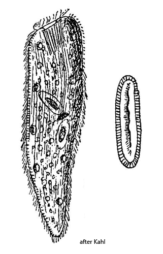
body elongated oval, tapering posteriorly from the middle
length 250–350 µm
mouth opening sligtly ventrally shifted
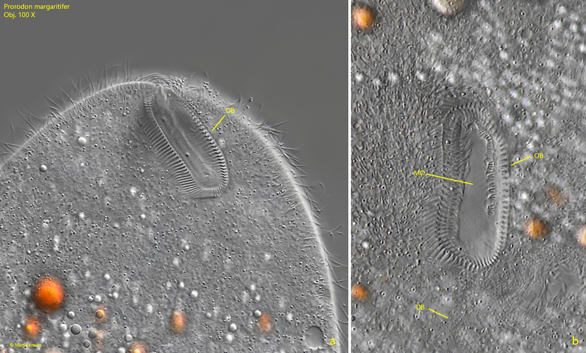
oral basket with > 80 rods, oval or almost rectangular
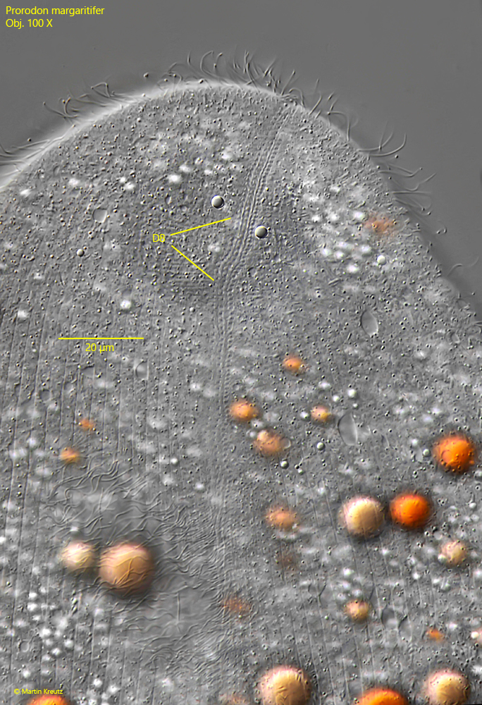
dorsal brush with 3 rows about one thrid of body length
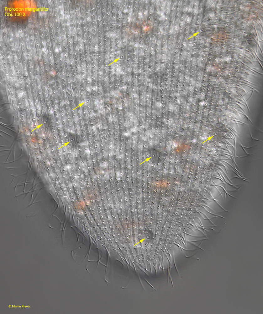
numerous contractile vacuoles scattered over body
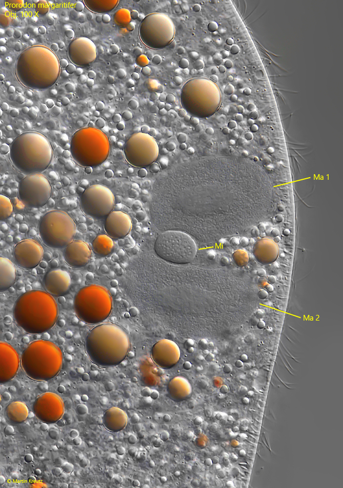
two ellipsoid macronuclei with oval micronucleus in between
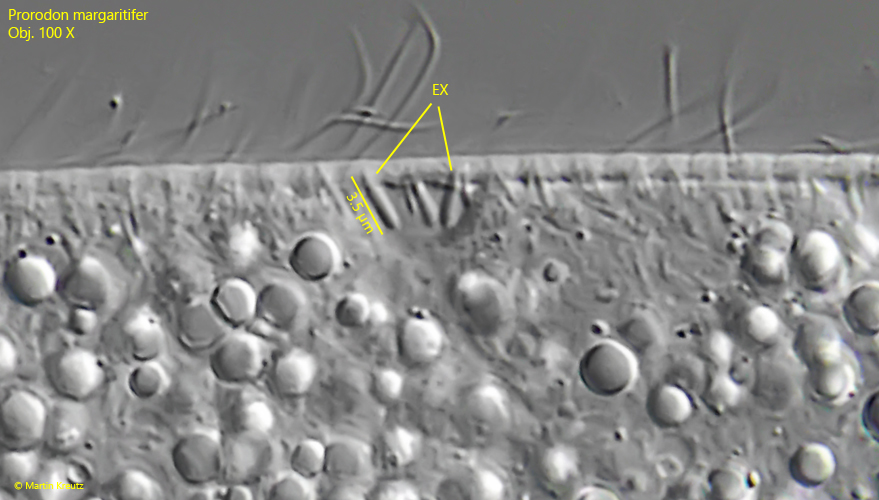
extrusomes club-shaped, about 3.5 µm long
Prorodon margaritifer
So far I have only found Prorodon margaritifer in the Simmelried and Bussenried. However, the species is quite rare in these locations. I find one specimen every 2 years on average.
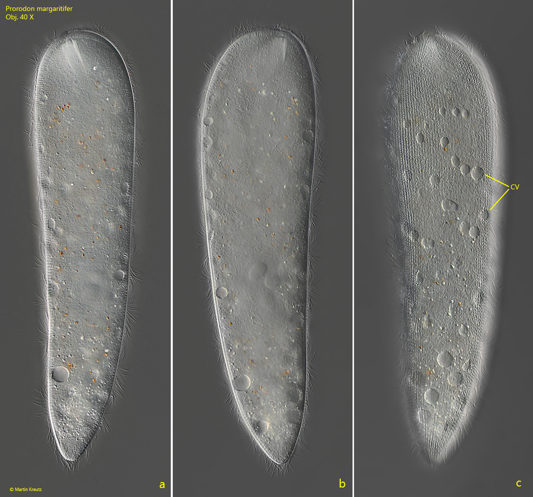
In a fresh sample, Prorodon margaritifer immediately stands out due to its size of about 300 µm and its rapid swimming movements. The ciliate often pushes back and forth. The mouth opening is slightly shifted ventrally and numerous contractile vacuoles are scattered over the body, which lie directly beneath the pellicular (s. fig. 1 c). The longitudinal rows of cilia are very dense. Even at medium magnification the nuclear apparatus can be recognized, which consists of two ellipsoidal macronuclei and an oval micronucleus between them (s. fig. 5). This is a clear distinguishing feature from Holophrya teres, which has a round macronucleus with an attached micronucleus.
The mouth opening of Prorodon margaritifer is long oval or almost rectangular (s. fig. 4 a-b). In my specimens the oral basket consisted of 82–86 rods. Kahl described the contractile vacuoles with 4 excretory pores each. In my population, however, there was always only one excretory pore in all contractile vacuoles (s. fig. 6). The extrusomes are small and inconspicuous. I was able to observe club-shaped ones with a length of about 3.5 µm (s. fig. 7). A second type of extrusomes seems to be present. These could be slightly curved rods of similar length. However, I was not able to recognize them exactly.
Fig. 1 a-c:Prorodon margaritifer. L = 325 µm. A freely swimming specimen. Note the numerous contractile vacuoles (CV) scattered over the body. Obj. 40 X.
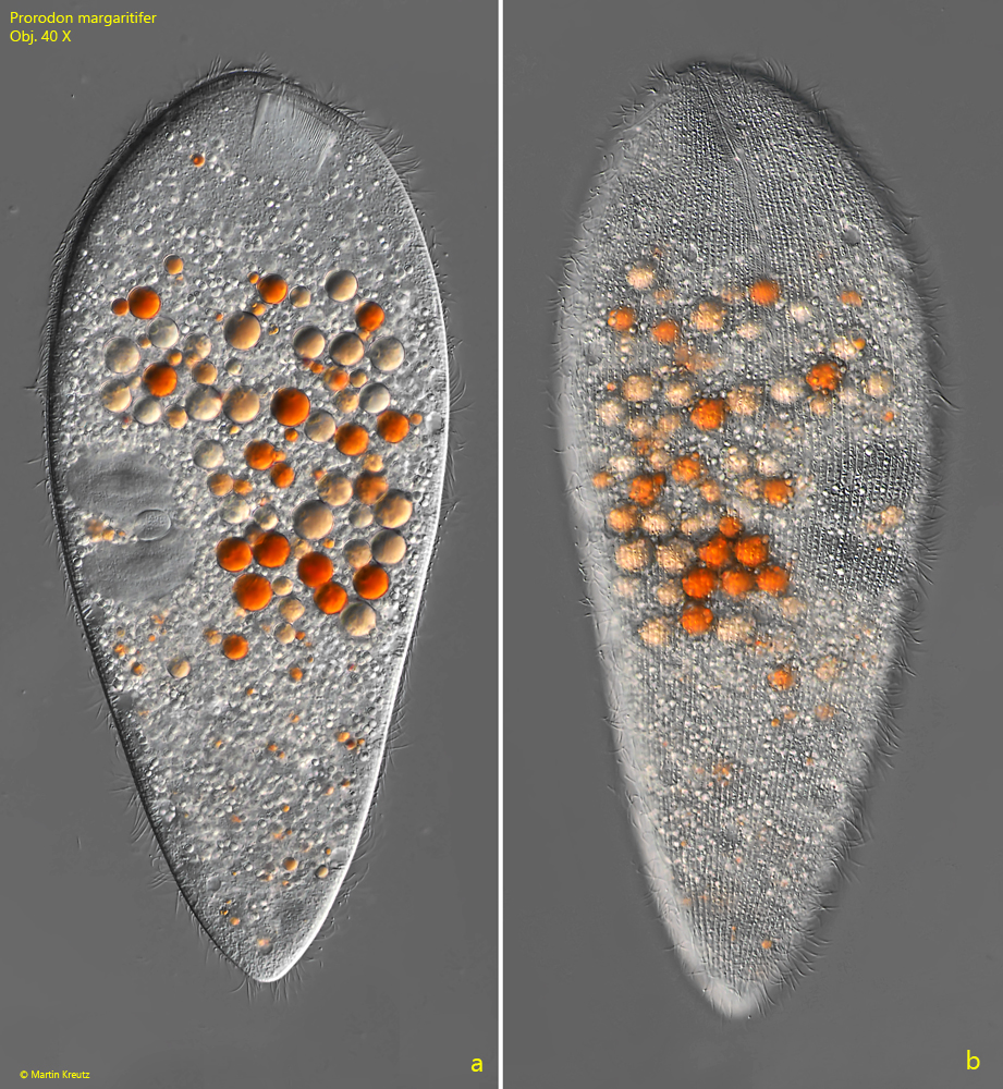
Fig. 2 a-b:Prorodon margaritifer. L = 310 µm. A second, slightly squashed specimen, coloured orange by oil droplets from ingested food. Obj. 40 X.
Fig. 3:Prorodon margaritifer. The three-rows dorsal brush (DB) with a length of about 125 µm in detail. Obj. 100 X.
Fig. 4 a-b:Prorodon margaritifer. The mouth opening (MO) is shaped elongated ovally or sometimes almost rectangular. The oral basket (OB) of the specimen is consisting of 85 rods. DB = part of the dorsal brush. Obj. 100 X.
Fig. 5:Prorodon margaritifer. The nuclear apparatus is consisting of two, macronuclei (Ma 1, Ma 2) and an oval shaped micronucleus (Mi) in between. Obj. 100 X.
Fig. 6:Prorodon margaritifer. The contractile vacuoles located beneath the pellicle have on excretion pore each (arrows). Obj. 100 X.
Fig. 7:Prorodon margaritifer. The club-shaped extrusomes are about 3.5 µm long. Obj. 100 X.