shell chitinoid, transparent, often brownish, 2 lateral pores (hard to see)
length 75–116 µm, width 33–64 µm
aperture with thickened lip
protoplast fill about half of shell, attached to shell with filaments of cytoplasm
lobose pseudopodia
several contractile vacuoles located in posterior half
nucleus spherical, diameter about 10 µm, with several nucleoli
Hyalosphenia elegans
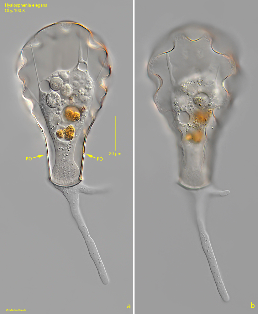
I found Hyalosphenia elegans in a small puddle in the Sima Moor (Austria) in June 2024. The conspicuous dents in the flask-shaped shell make it easy to identify the species. The pseudopodia are lobose and are only extended by undisturbed specimens. The protoplast of the amoeba does not completely fill the shell and is attached to the posterior part of the shell with filaments of cytoplasm (s. figs. 2 a and 3). According to my observations, Hyalosphenia elegans has several contractile vacuoles, which are localized in the posterior half of the body. The spherical nucleus is also located posteriorly and has a diameter of about 10 µm. Several spherical nucleoli of different size are visible in the nucleus.
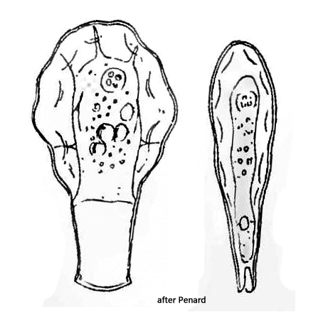
Penard (1902) already described 2 lateral pores, which are located at the neck of the shell. They can only be identified at high magnification (s. figs. 2 a and 4). The function of these pores is unknown.
Fig. 1 a-d:Hyalosphenia elegans. L = 92 µm (of shell). Different focal planes of an unsquashed specimen. Obj. 60 X.
Fig. 2 a-b:Hyalosphenia elegans. L = 92 µm (of shell). The same specimen as shown in fig. 1 a-d in detail. PO = lateral pores of the neck. Obj. 100 X.
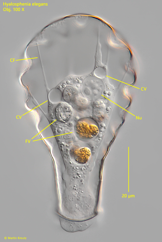
Fig. 3:Hyalosphenia elegans. L = 92 µm (of shell). The slightly squashed specimen as shown in fig. 2 a-b. CF = filaments of cytoplasm attached to the shell, CV = contractile vacuoles, FV = food vacuoles, Nu = nucleus. Obj. 100 X.
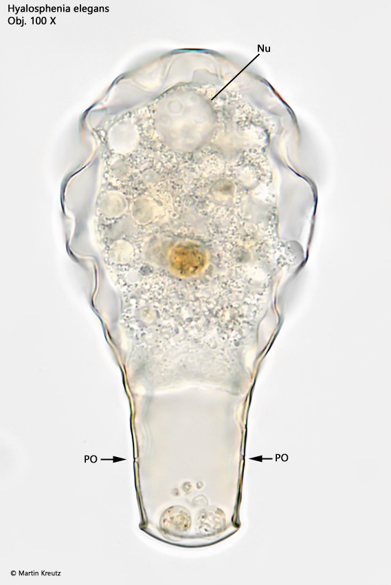
Fig. 4:Hyalosphenia elegans. L = 89 µm (of shell). Focal plane on the two pores (PO) located at the lateral sides of the neck. Nu = nucleus. Obj. 100 X.