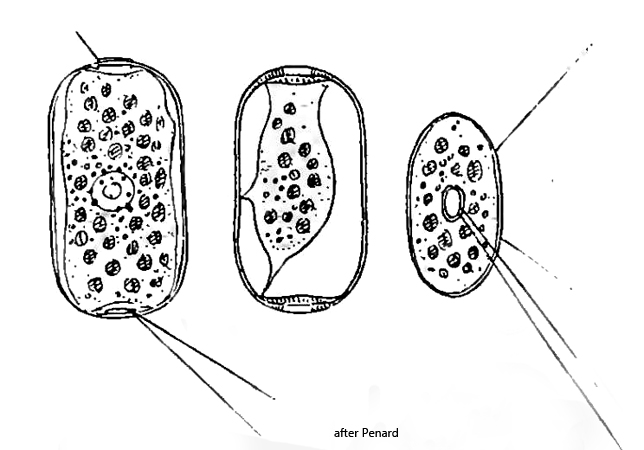
shell ovoid to elliptical, compressed, brownish transparent
shell with two elliptical apertures at opposite ends
length 45–77 µm
several contractile vacuoles
nucleus spherical with central nucleolus
filopodia thin, straight, sometimes branched
symbiotic algae present
Archerella flavum
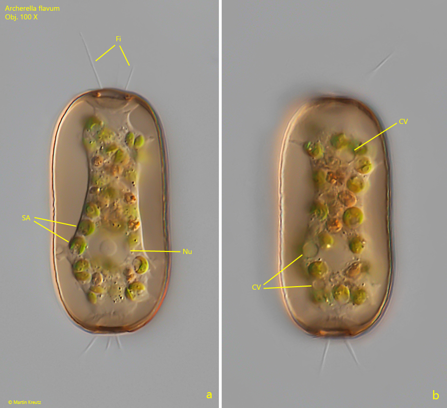
I found Archerella flavum in a small puddle in the Sima Moor (Austria) in June 2024. This filose testate amoeba is easy to identify by its conspicuous shell. It is usually brownish in color, transparent, without attached xenosomes and has two opposite apertures. The protoplast can be seen clearly through the transparent shell. It is attached to the inner wall of the shell with strands of cytoplasm. The thin, mostly straight filopodia are only extended by undisturbed specimens (s. figs. 1 a and 2 a).
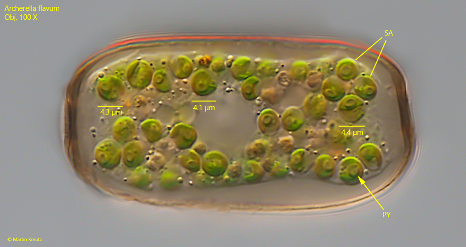
The protoplast of Archerella flavum is usually clearly green in color because this testate amoeba has symbiotic algae. These algae have never been described in detail in the literature available to me. According to my observations, the algae cells are spherical with a diameter of 4.0–4.5 µm. They seem to be of the Chlorella type. They have a cup-shaped chloroplast, each with a pyrenoid. I could also see that each algae cell has a separate nucleus.
Fig. 1 a-b:Archerella flavum. L = 50 µm. Two focal planes of an unsquashed specimen. Note the filopodia (Fi), which are extended from the two apertures of the shell. CV = contractile vacuoles, Nu = nucleus, SA = symbiotic algae. Obj. 100 X.
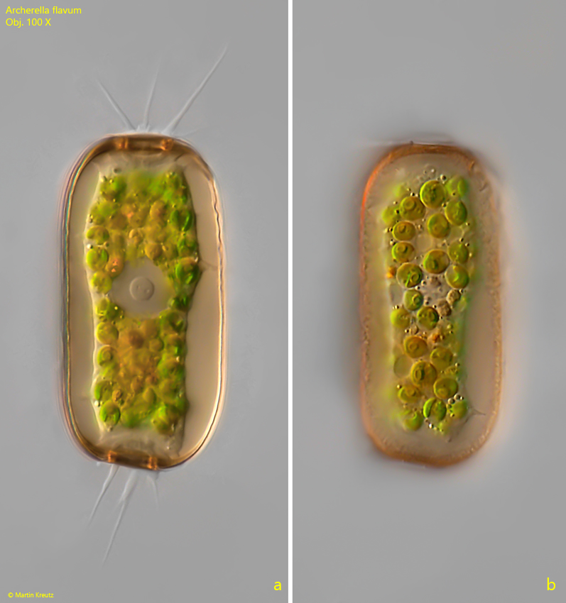
Fig. 2 a-b:Archerella flavum. L = 53 µm. Two focal planes of a second specimen with extended filopodia. Obj. 100 X.
Fig. 3:Archerella flavum. L = 53 µm. The slightly squashed specimen as shown in fig. 2 a-b to show the symbiotic algae (SA) in detail. The algae seem to be of the Chlorella type with a cup-shaped chloroplast with a single pyrenoid (PY). Obj. 100 X.