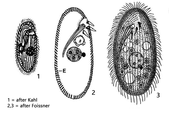
Without nassulid frange of membranelles oral aperture as in Nassula
three membranelles on the left of oral aperture
plasm colorless or slightly reddish/orange
lateral extrusomes are directed to the posterior end
the pharyngael rods do not reach the surface
Furgasonia theresae
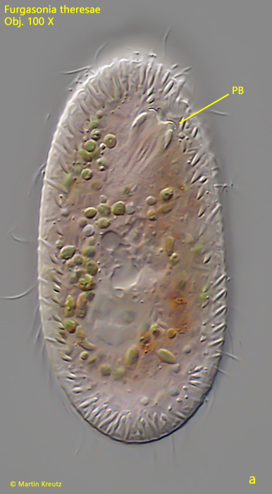
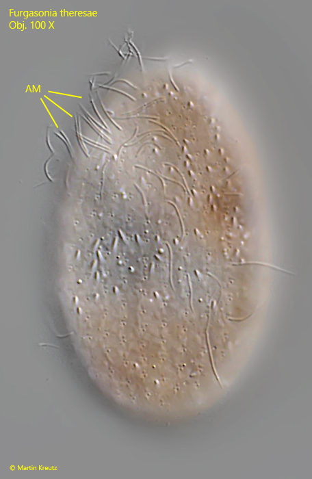
I have found Furgasonia theresae exclusively in Simmelried in floating, decaying plants. The distinction from Nassula is best made on the basis of the backward directed extrusomes, and the sunken pharyngeal basket, which does not reach the surface (s. fig. 2 a). The population in Simmelried also shows a reddish-orange coloration as already described by Kahl (as Cyclogramma trichocystis). Furthermore, Furgasonia theresae is with 70 µm smaller than most representatives of the genus Nassula. The adoral membranelles of Furgasonia, which are located to the left of the mouth opening, are difficult to see light microscopically (s. fig. 3).
The history of the taxonomic classification of Furgasonia theresae is interesting and complex. The species was first referred to as Nassula theresae by Fabre-Domergue (1889). Five years later it was published independently of Fabre-Domergue as Nassula trichocystis by Stokes (1895). Kahl, as the first reviser of the group, could not obtain the publication of Fabre-Domergue and treated Nassula trichocystis as Cyclogramma (Nassula) trichocystis (Kahl, 1931). The genus Cyclogramma was later declared as invalid, due to the lack of a genus diagnosis. Then in 1989 by Foissner Nassula theresae and Nassula trichocystis were synonymized to Nassula theresae (because of the earlier publication by Fabre-Domergue) and transferred into the genus Furgasonia, defined before by Jankowski (1964), so that the species is called Furgasonia theresae today.
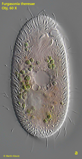
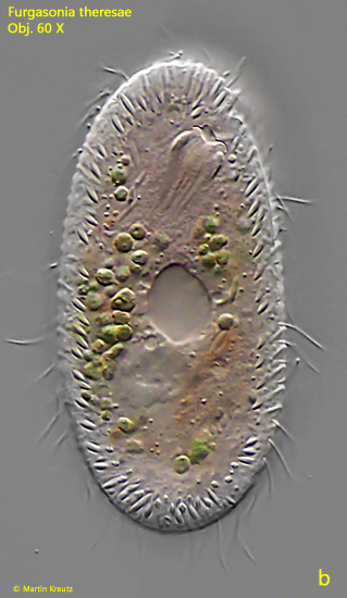
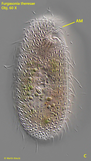
Fig. 1 a-c:Furgasonia theresae. L = 70 µm. Three focal planes of a freely swimming specimen. AM = adoral membranelle. Obj. 60 X.
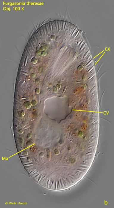
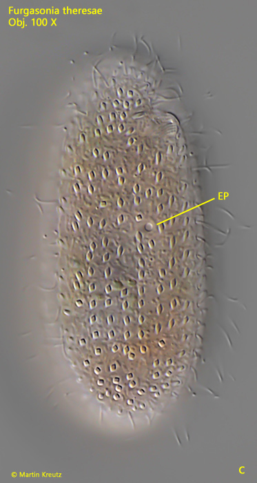
Fig. 2 a-c:Furgasonia theresae. L = 70 µm. Three focal planes of a slightly squashed specimen. CV = contractile vacuole, EP = excretion porus, EX = extrusomes, Ma = macronucleus, PB = pharyngeal basket. Obj. 100 X.
Fig. 3:Furgasonia theresae. The adoral membranelles (AM) at the left side of the mouth opening in a squashed specimen. Obj. 100 X.
Fig. 4:Furgasonia theresae. L = 55 µm. Focal plane on the macronucleus (Ma) and micronucleus (Mi) in a slightly squashed specimen. EX = extrusomes, PB = pharyngeal basket. Obj. 100 X.