body elongate ellipsoid, almost parallel sides, laterally flattened
length 200–220 µm, width 30–40 µm
oral bulge indistinct, discoid
type 1 extrusomes 5–6 µm long curved rods
type 2 extrusomes straight and thin, 30–40 µm long
cortex covered with mucilaginous layer
macronucleus kidney-shaped, sometimes elongated or three-lobed
micronucleus located in concave indentation of macronucleus
contractile vacuole terminal
Enchelys vestita
I regularly find Enchelys vestita in the Simmelried. I usually find the specimens in the uppermost layer of mud. In January 2000 I was able to observe a mass development, with about 10 specimens per milliliter. Many specimens of Ileonema simplex were also found in the same sample.
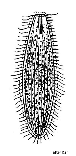
The only available description and drawing of Enchelys vestita is by Kahl (1930). In my opinion, his drawing is not very informative, because he drew the body quite compact and because he describes and draws the macronucleus as elongated oval. In fact, the macronucleus in this species is subject to a certain amount of variation, but it is usually clearly kidney-shaped (s. figs. 1 a, 6 and 7). Sometimes the macronucleus is also elongated C-shaped or trilobed (s. fig. 2 b), but the ends are always thickened. Another important chracteristic is the mucilaginous layer covering the pellicle. It appears granulated and sometimes “scaly” due to the embedded lepidosomes (s. fig. 5).
Kahl gives a length of about 25 µm for the extrusomes. However, I was able to observe that there are at least two types of extrusomes arranged in the oral bulge of Enchelys vestita. The shorter ones are 5–6 µm long rods which are slightly curved. The longer extrusomes (which Kahl probably meant) are straight and thin rods, but according to my measurements they are 30–40 µm long (s. fig. 3).
In many specimens of Enchelys vestita I observed coccal algae in the food vacuoles (s. figs. 4 and 7). Since all algae were of the same type, it is possible that ciliates with symbiotic algae are among the prey organisms of Enchelys vestita.
Enchelys vestita is only slightly contractile. The contraction is also not as fast as in the genus Spirostomum, for example. The maximum contraction is about 20 % of the body length.
When I had the opportunity to measure several specimens in January 2000, I recorded some morphometric data of Enchelys vestita (s. tabl. 1).
Table 1: Morphometric data an Enchelys vestita.
Characteristics
n
mean
min
max
body length (µm)
30
194
135
276
body width (µm)
30
38
18
65
macronucleus length (µm)
20
38
25
65
macronucleus width (µm)
20
16
11
26
Number of micronuclei
14
1
1
1
diameter micronucleus (µm)
13
4.8
4
7.2
thickness of mucilaginous layer (µm)
26
2.2
1.8
2.7
In the year 2016 Foissner described the new species Cataphractes austriacus found in a small Sphagnum bog near Salzburg (Austria). He assumed, that Cataphactes austriacus could be synonym with Enchelys vestita. However, according my observations of Enchelys vestita this is not the case. The length of Cataphractes austriacus is on average 120 µm long while the lengths of Enchelys vestita is 200 µm. The body of Enchelys vestita ist distinctly flattened (s. fig. 4) and the macronucleus is kidney-shaped, what is not the case for Cataphractes austriacus (body not flattened, macronucleus elongated oval). Finally, in Cataphractes austriacus the curved type 1 extrusomes are only 2–3 µm long and the acicular type 2 extrusomes are 25 µm long. This set of extrusomes are not identical to the extrusomes present in Enchelys vestita.
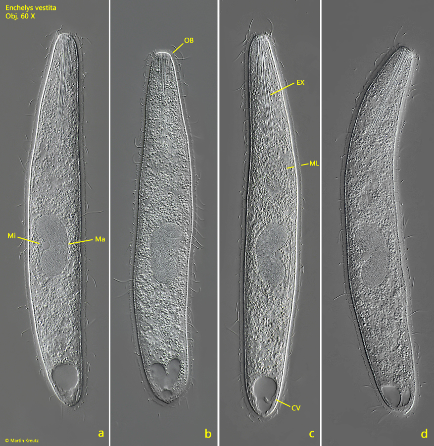
Fig. 1 a-d:Enchelys vestita. L = 223 µm. A freely swimming specimen. Note the kidney-shaped macronucleus (Ma) and the mucilaginous layer (ML) covering the cortex. CV = contractile vacuole, EX = extrusomes, Mi = micronucleus, OB = oral bulge. Obj. 60 X.
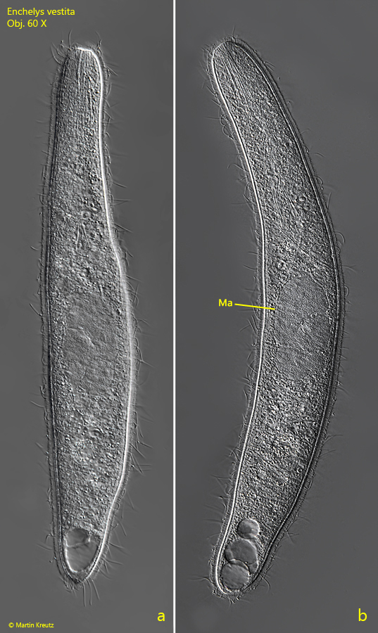
Fig. 2 a-b:Enchelys vestita. L = 275 µm. A second, freely swimming specimen with a three-lobed macronucleus (Ma). Obj. 60 X.
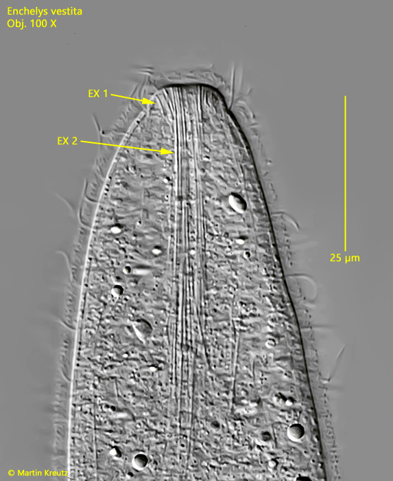
Fig. 3:Enchelys vestita. The oral bulge is armed with two types of extrusomes. Type 1 are 5–6 µm long and slightly curved (EX 1), while type 2 are straight and thin rods with a length of 30–40 µm (EX 2). Obj. 100 X.
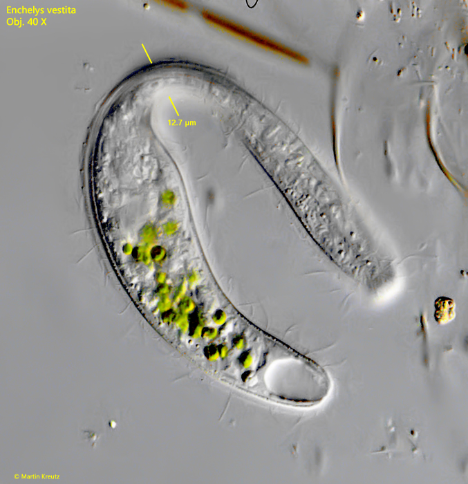
Fig. 4:Enchelys vestita. L = 188 µm. In a rotating specimen the lateral flattening of the body is visible. The specimen is 12.7 µm thick. Obj. 60 X.
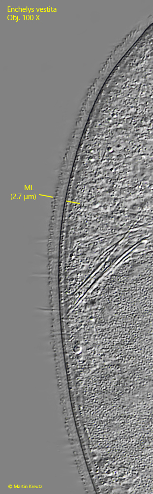
Fig. 5:Enchelys vestita. The mucilaginous layer (ML) of a squashed specimen in detail. The layer appears granulated due to the scattered lepidosomes in the layer which are below the resolution limit. Obj. 100 X.
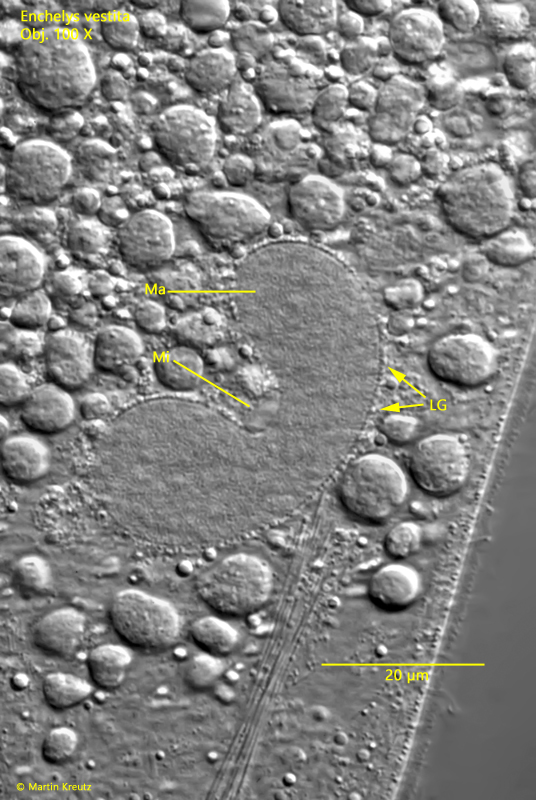
Fig. 6:Enchelys vestita. The kidney-shaped macronucleus (Ma) is often covered with a thin layer of granules (LG). Mi = micronucleus. Obj. 100 X.
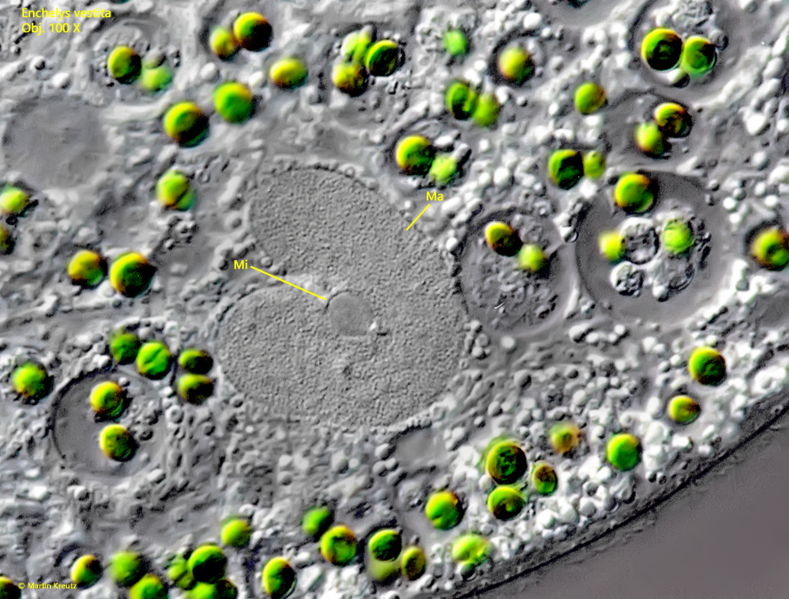
Fig. 7:Enchelys vestita. The macronucleus (Ma) and the micronucleus (Mi) of a second specimen. Obj. 100 X.