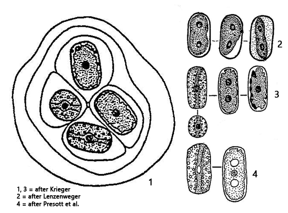
cells cylindrical, sometimes ovoid, apices broadly rounded
length 10–44 µm, width 5–20 µm
one chloroplast plate shaped, with wavy margin
1–2 central pyrenoids, covered with starch grains
cells embedded in layered gelatinous mass
nucleus central
cytoplasm sometimes colored slightly violet
Mesotaenium macrococcum
I have only found Mesotaenium macrococcum once in samples from the Schwemm Moor in Austria. According to Lenzenweger (2003), the species should also occur on moist soils, rocks and between mosses.
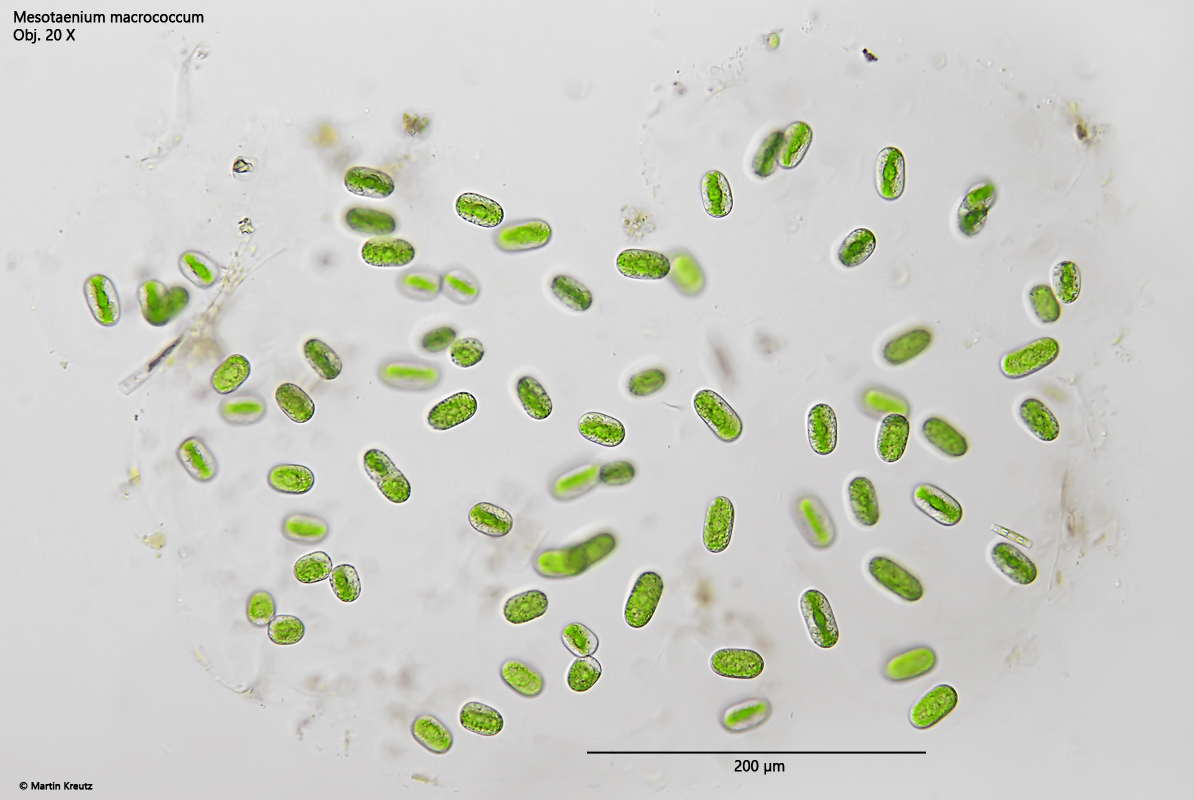
The samples from the Schwemm Moor contained colonies with approx. 10–30 cells in a indistinctly layered gelatinous matrix. The cells were quite widely separated from each other in the matrix (s. fig. 1). The most important characteristic of Mesotaenium macrococcum is the plate-shaped chloroplast. (s. figs 4 a-b and 5 a-b). The cells of my population were between 20–36 µm long. This means that the largest cells were significantly longer than in the variety Mesotaenium macrococcum var. minus with 19–26 µm. In the second similar species Mesotaenium chlamydosporum with cells of comparable length (15–33 µm), the plate-shaped chloroplast is clearly divided into two parts in larger cells, which was not the case in my species.
Fig. 1:Mesotaenium macrococcum. D = 600 µm (of colony). A slightly squashed colony in a layered gelatinous mass. Obj. 20 X.
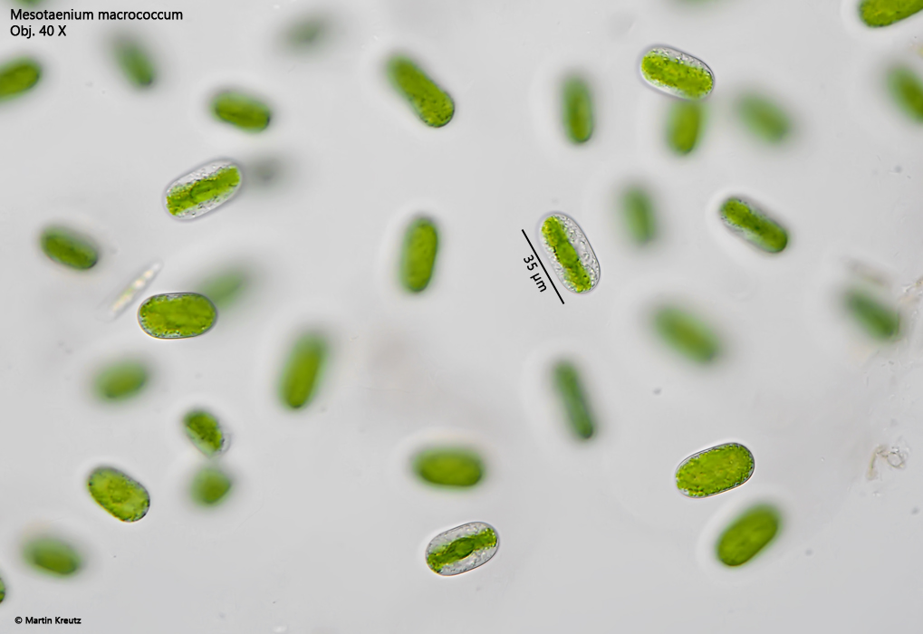
Fig. 2:Mesotaenium macrococcum. L = 21–35 µm (of cells). Some cells of the colony as showed in fig. 1. Obj. 40 X.
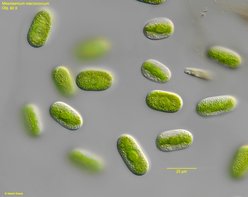
Fig. 3:Mesotaenium macrococcum. L = 18–34 µm. Some cells with a plate-shaped chloroplast in frontal view and in lateral view. Obj. 60 X.
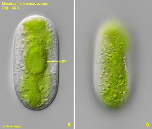
Fig. 4 a-b:Mesotaenium macrococcum. L = 36 µm. A cell with the plate-shaped chloroplast in lateral view. The pyrenoid (PY) is located in the center of the chloroplast. Obj. 100 X.
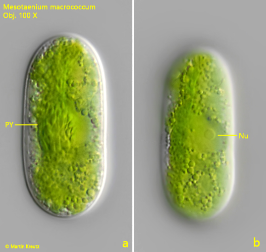
Fig. 5 a-b:Mesotaenium macrococcum. L = 38 µm. A cell with frontal view on the plate-shaped chloroplast. The pyrenoid (PY) is covered with tightly packed starch grains. The nucleus (Nu) is located adjacent to the chloroplast. Obj. 100 X.
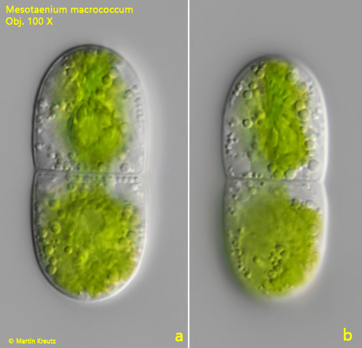
Fig. 6 a-b:Mesotaenium macrococcum. L = 38 µm. A specimen during cell division. Obj. 100 X.