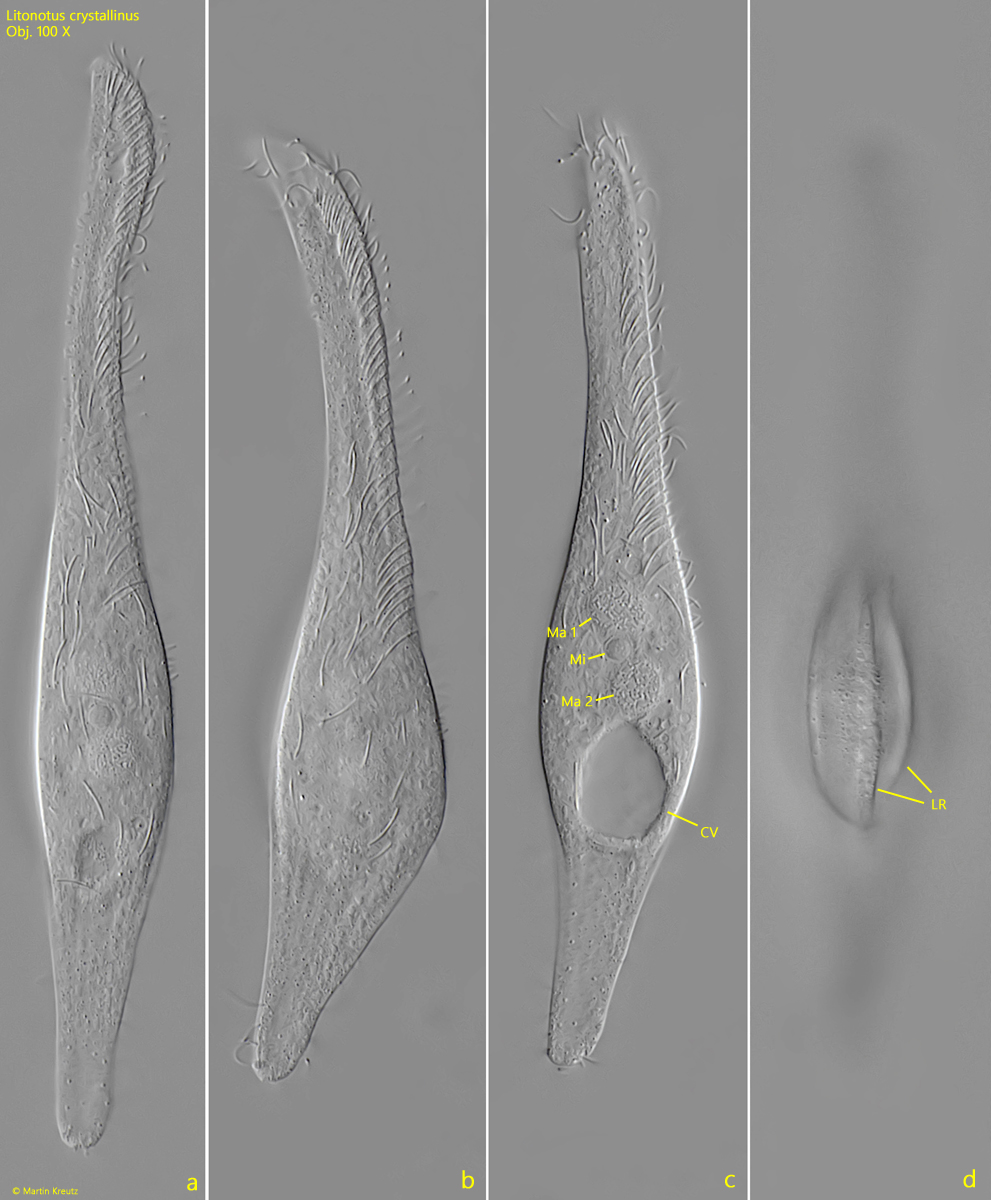
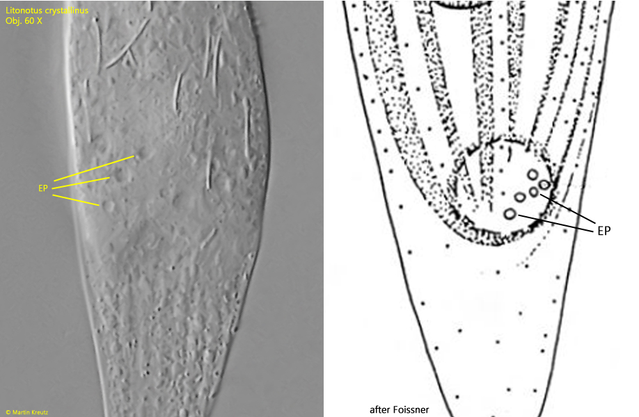
Litonotus crystallinus
(Vuxanovici, 1960) Foissner, Berger, Blatterer & Kohmann, 1995
Most likely ID: n.a.
Synonym: Lionotus crystallinus
Sampling location: Mühlhalden pond, Simmelried
Phylogenetic tree: Litonotus crystallinus
Diagnosis:
- body slenderly to broadly lanceolate, laterally flattened
- anterior end bent dorsally and pointed, ventral side convex
- posterior end narrowly rounded or tail-like
- length 80–188 µm
- left side with 3–5 distinct ribs
- right side with 5–8 rows of cilia
- dorsal brush of club-shaped, short cilia
- two spherical macronuclei
- one spherical micronucleus between the macronuclei
- extrusomes rod-shaped, slightly curved, about µm long
- extrusomes arranged mainly along oral cleft
- contractile vacuole clearly subterminal
- rarely a second contractile vacuole in anterior third
I find Litonotus crystallinus only rarely. In fresh samples, the specimens are difficult to find because they often adhere to detritus flakes. However, sometimes specimens can be found that have settled on the floating coverslip. These specimens can then only be examined from the right, ciliated side, with which they glide on the coverslip.
My specimens were between 150–170 µm long. I could only recognize the characteristic ribs on the left side of the body by focusing through from the right side (s. fig. 2 d). According to Foissner et al. (1995), the extrusomes are described as slightly curved with a length of about 6 µm. In my specimens, the extrusomes were indeed curved but measured 8.0–8.7 µm in length (s. fig. 5). Apart from the data from Foissner et al., I have found no other study on the extrusomes in Litonotus crystallinus. Therefore, it is unclear how much variability there can be in the extrusomes of this species.
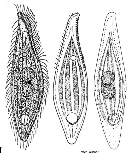
The distinction from Litonotus cygnus is difficult. Large specimens of Litonotus crystallinus can be as long as smaller, or contracted, Litonotus cygnus. The extrusomes of Litonotus cygnus are also curved and as large as those of Litonotus cygnus. I consider the ribs on the left side of Litonotus crystallinus to be the most reliable distinguishing feature.
Fig. 1 a-c: Litonotus crystallinus. L = 166 µm. A freely swimming specimen from right. Obj. 60 X.
Fig. 2 a-d: Litonotus crystallinus. L = 166 µm. The same specimen as shown in fig. 1 a-c from right. Note the ribs on the left side (LR) focused through from the right side. The micronucleus (Mi) is located between two spherical macronuclei (Ma 1, Ma 2). CV = contractile vacuole. Obj. 100 X.
Fig. 3: Litonotus crystallinus. A part of the dorsal brush (DB) with pairs of club-shaped, short cilia (arrows). Obj. 100 X.
Fig. 4: Litonotus crystallinus. There are several excretions pores (EP) of the contractile vacuole on the left side (focused through from the right side) as drawn bei Foissner. Obj. 100 X.
Fig. 5: Litonotus crystallinus. The slightly curved extrusomes (EX) in the squashed specimen have a length of 8.0–8.7 µm. Ma 1 + Ma 2 = macronuclei, Mi = micronucleus. Obj. 100 X.