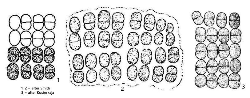
colonies flat, rectancular, cells in rectangular rows
cells oval or ellipsoid, rarely spherical, 5–9 X 4–7 µm
cells during division hemispherical
gelatinous sheat not firm, about 10 µm thick
cytoplasm with granules
color blueish-green or greenish
Merismopedia elegans
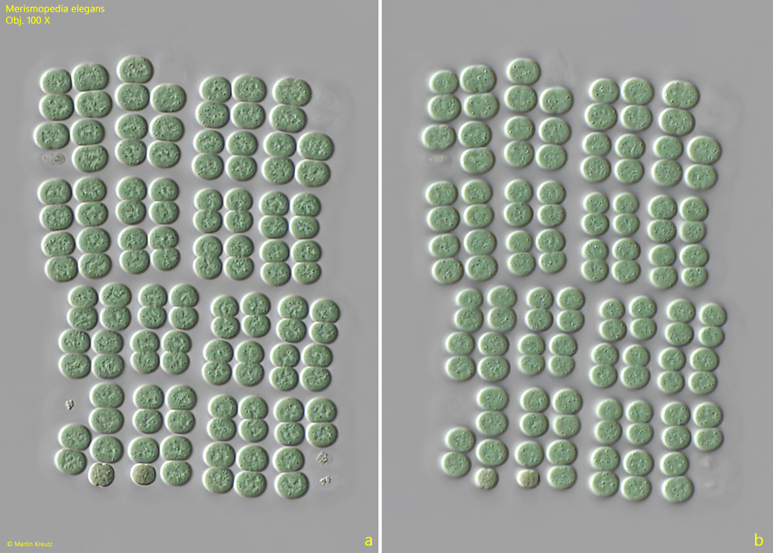
I find Merismopedia elegans only very rarely in the Simmelried. In the Schwemm Moor (Austria), this cyanobacterium was much more frequently found.
The colonies of Merismopedia elegans can become very large because the cells lie in a common gelatinous sheath, which does not break down even after several cell divisions. The gelatinous sheath extends about 10 µm beyond the colonies and can be sharply or indistinctly bounded. Smaller colonies are flat and plate-shaped, while larger ones can also be wavy and curved.
Merismopedia elegans can be recognized by the comparatively large cells, which are usually longer than 5 µm. The cells of most other species within the genus Merismopedia are significantly smaller. In addition, the cytoplasm of the cells contains clearly visible granules. In my population, the cells were 6–8 µm long and about 5-6 µm wide. The granules in the cytoplasm were more concentrated in the center of the cells. The colonies were all blue-green in varying intensities.
Fig. 1 a-b:Merismopedia elegans. L = 92 µm (of colony). Two focal planes of a colony of 128 cells. Obj. 100 X.
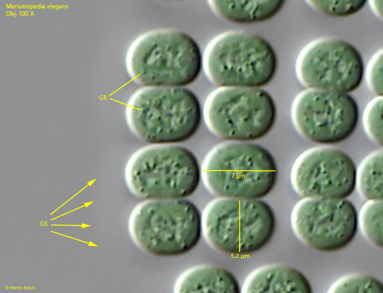
Fig. 2:Merismopedia elegans. L = 7 µm (of cells). A crop from fig. 1 a. The colony is embedded in a gelatinous sheath (GS). In the cytoplasm of the cells granules (GR) are scattered. Obj. 100 X.
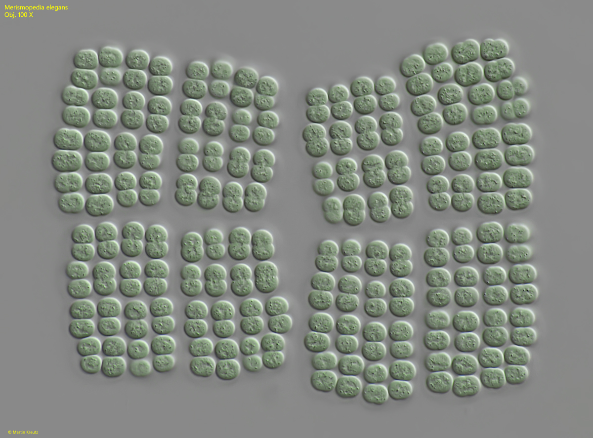
Fig. 3:Merismopedia elegans. L = 130 µm (of colony). A second colony of 256 cells. Obj. 100 X.
Fig. 4 a-b:Merismopedia elegans. L = 75 µm. A third colony of 128 cells in DIC (a) and in brightfield illumination (b). Obj. 100 X.