zooids almost fusiform, dorsal side more convex than ventral
aggregated in pseudocolonies of < 10 zooids
contracted zooids almost spherical
length 28–92 µm
peristome slightly oblique
macronucleus horseshoe-shaped in anterior half
one micronucleus, 3–4 µm
one contractile vacuole on dorsal side
pellicle with delicate, narrow striation
stalk thin, 1.5–2 µm in diameter, branched or unbranched
Opercularia asymmetrica
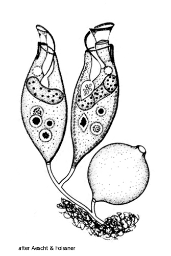
Opercularia asymmetrica was first described by Biczok (1956) as Pyxidium asymmetricum. Some years later Guhl (1979) synonymized Pyxidium asymmetricum with Opercularia coarctata to Opercularia asymmetrica. In 1992, Aecht & Foissner published a redescription of Opercularia asymmetrica, concluding that Opercularia asymmetrica is a valid, independent species, which is not synonymous with Opercularia coarctata.
Like Biczok, I found Opercularia asymmetrica in a hay infusion, in which the species developed in large numbers. The pseudocolonies grew mainly on bacterial flocs near the surface but also on the vessel wall. While Biczok as well as Aescht & Foissner mainly found solitary and stalkless zooids, in my case, there were mainly small pseudocolonies of 4–6 zooids on thin, branched stalks (s. fig. 1 and 2).
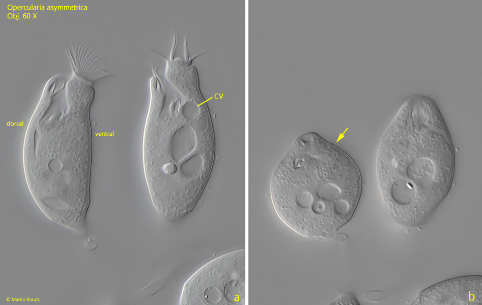
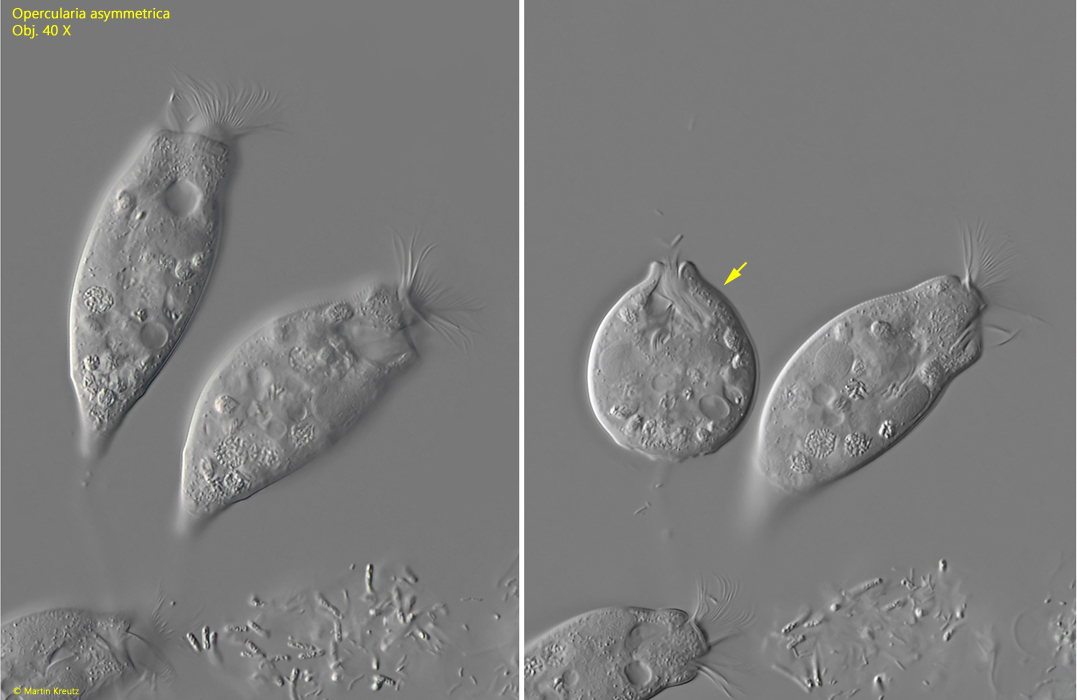
The zooids of my population were between 40–95 µm long and had a slightly asymmetrical body shape. The dorsal side is more strongly convexly curved than the ventral side (s. fig. 3 a). On the dorsal side, the contractile vacuole can be easily seen, which is clearly distant from the apical end (s. fig. 3 a). The contracted zooids were almost spherical and about half as long as the extended zooids (s. figs. 3 a-b and 4 a-b).
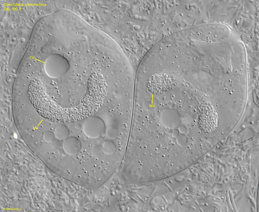
The stalks of unsquashed specimens were 2.5–3.0 µm thick and only branched into short offshoots at the distal end. In strongly squashed zooids, it was noticeable that the stalk has a distinct longitudinal striation (s. fig. 10). The macronucleus is horseshoe-shaped and lies transversely in the front half of the cell (s. fig. 7). I was able to identify a round micronucleus (s. fig. 8). The very delicate and tight striation of the pellicle can only be seen in slightly compressed specimens at high magnification (s. fig. 9).
Opercularia asymmetrica differs from the similar species Opercularia coarctata mainly by the position of the contractile vacuole. In Opercularia coarctata, it is located on the ventral side of the oral funnel. Additionally, the pellicle of Opercularia coarctata is smooth without transverse striation, and the stalks do not have longitudinal striation. With a maximum of 65 µm, Opercularia coarctata is also somewhat smaller than Opercularia asymmetrica.
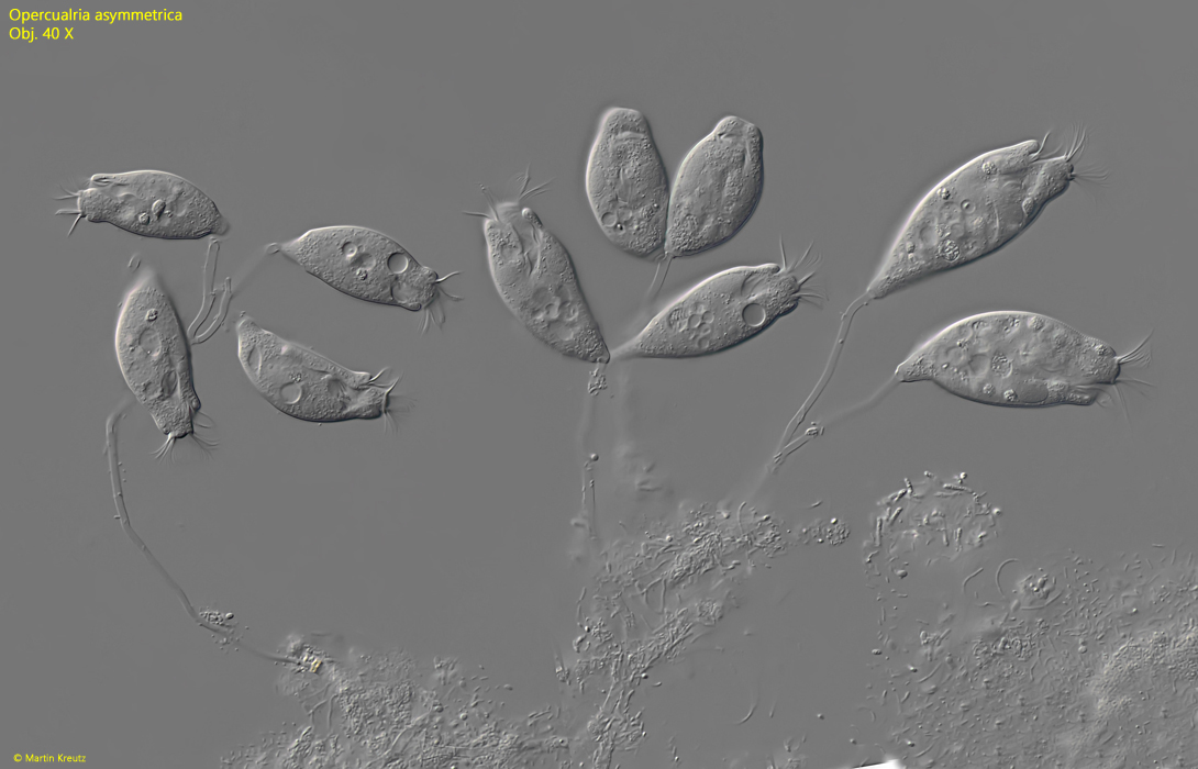
Fig. 1:Opercularia asymmetrica. L = 57–91 µm. Two pseudocolonies on thin, branches stalks. Obj. 40 X.
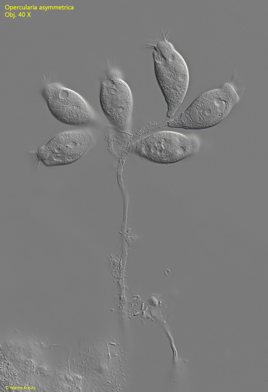
Fig. 2:Opercularia asymmetrica. L = 45–57 µm. A second pseudocolony of six zooids on a branched stalk. Obj. 40 X.
Fig. 3 a-b:Opercularia asymmetrica. L = 65 µm. An elongated and retracted specimen (arrow). Note the contractile vacuole (CV) on the more convex dorsal side. Obj. 60 X.
Fig. 4 a-b:Opercularia asymmetrica. L = 95 µm. A second elongated and retracted specimen (arrow). Obj. 40 X.
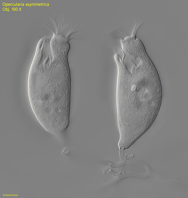
Fig. 5:Opercularia asymmetrica. L = 64–68 µm. Two elongated zooids. Obj. 100 X.
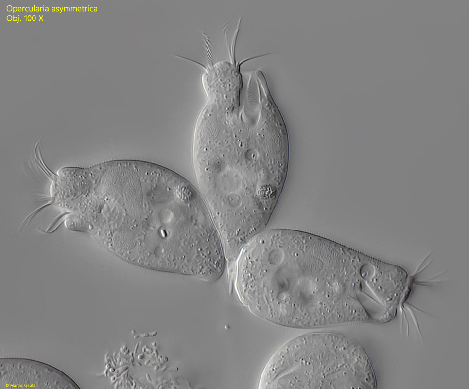
Fig. 6:Opercularia asymmetrica. L = 58–63 µm. Three elongated zooids. Obj. 100 X.
Fig. 7:Opercularia asymmetrica. The horseshoe-shaped macronucleus (Ma) in two squashed zooids. CV = contractile vacuole. Obj. 100 X.
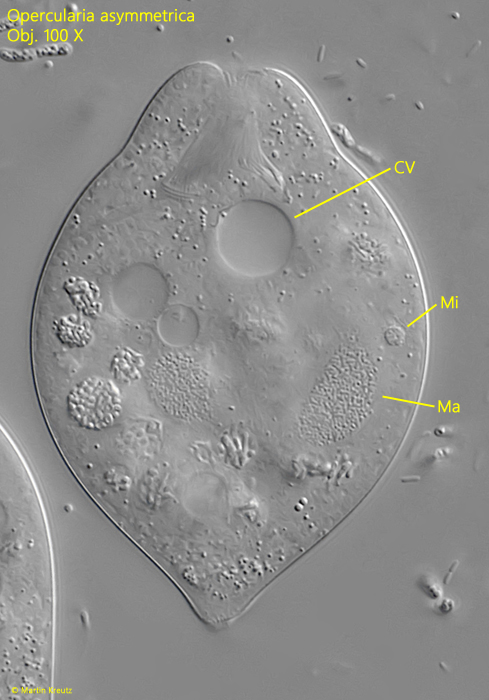
Fig. 8:Opercularia asymmetrica. The macronucleus (Ma) and the micronucleus (Mi) in a second, squashed zooid. CV = contractile vacuole. Obj. 100 X.
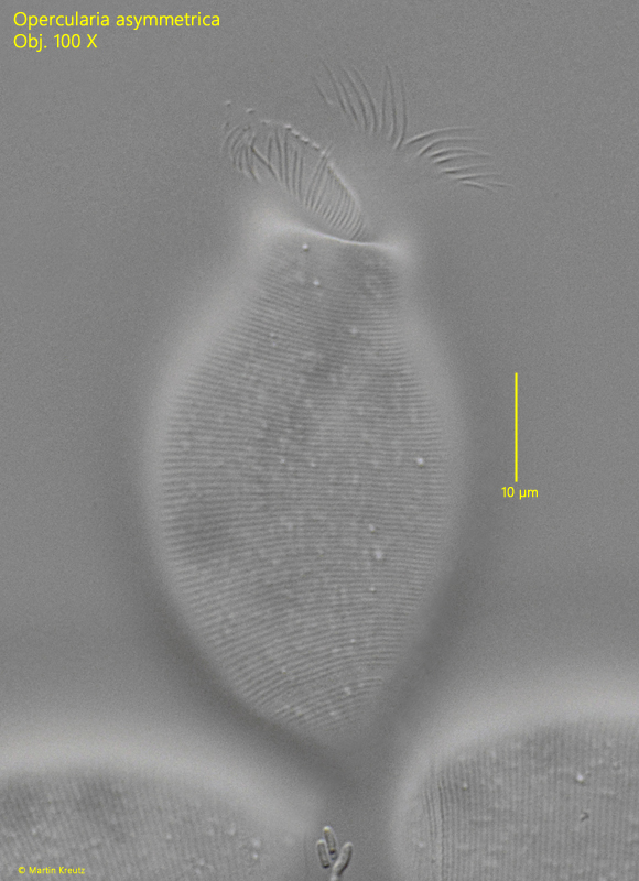
Fig. 9:Opercularia asymmetrica. The delicate striation of the pellicle in a slightly squashed zooid. Obj. 100 X.
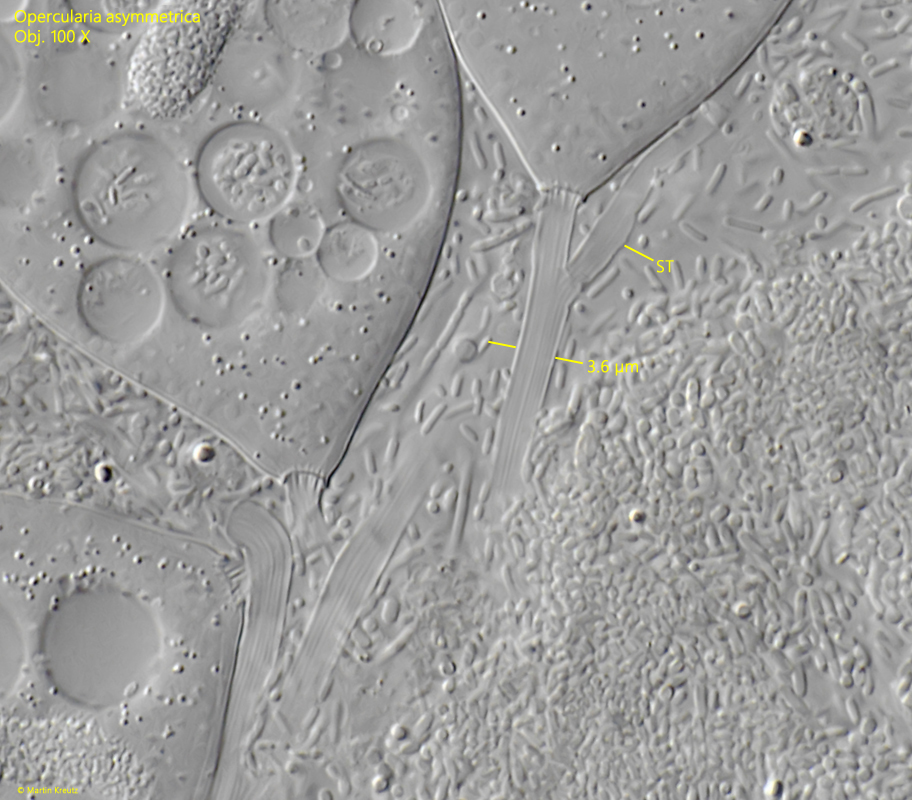
Fig. 10:Opercularia asymmetrica. The stalk (ST) in this strongly squashed pseudocolony has a diameter of 3.6 µm with a longitudinal striation. Obj. 100 X.