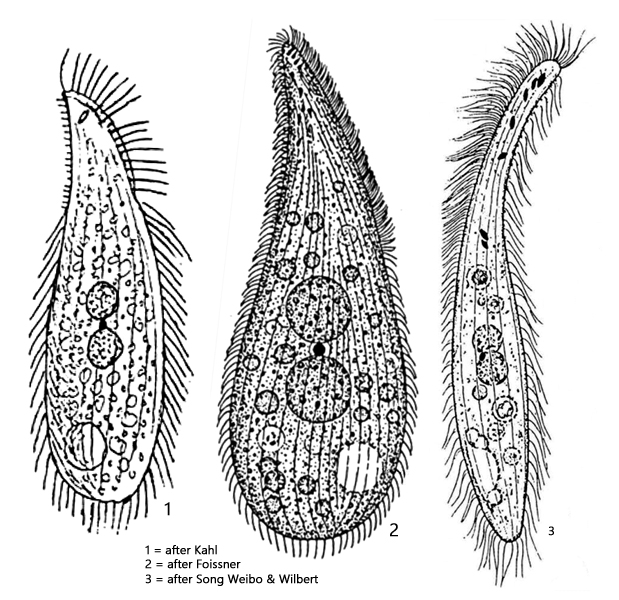
body slender to plump lanceolate, laterally flattened
length 80–150 µm
right side with 18–25 longitudinal rows of cilia
left side almost naked apart from 4–6 rows of short brisles
two ellipsoid or spherical macronuclei
one micronucleus between the macronuclei
extrusomes drop-shaped, about 3×1.5 µm
contractile vacuole sub-terminal on ventral side
Amphileptus punctatus
I find Amphileptus punctatus very rarely and always only single specimens. Almost all findings I made in autumn among decomposing leaves at the bottom of the waters.
The most striking feature of Amphileptus punctatus are the drop-shaped extrusomes, which are highly refractive and therefore noticeable even at small magnifications (s. figs. 2 a, 4 and 6). The body is slightly sigmoid-shaped, and in my population, the specimens were between 130–175 µm long.
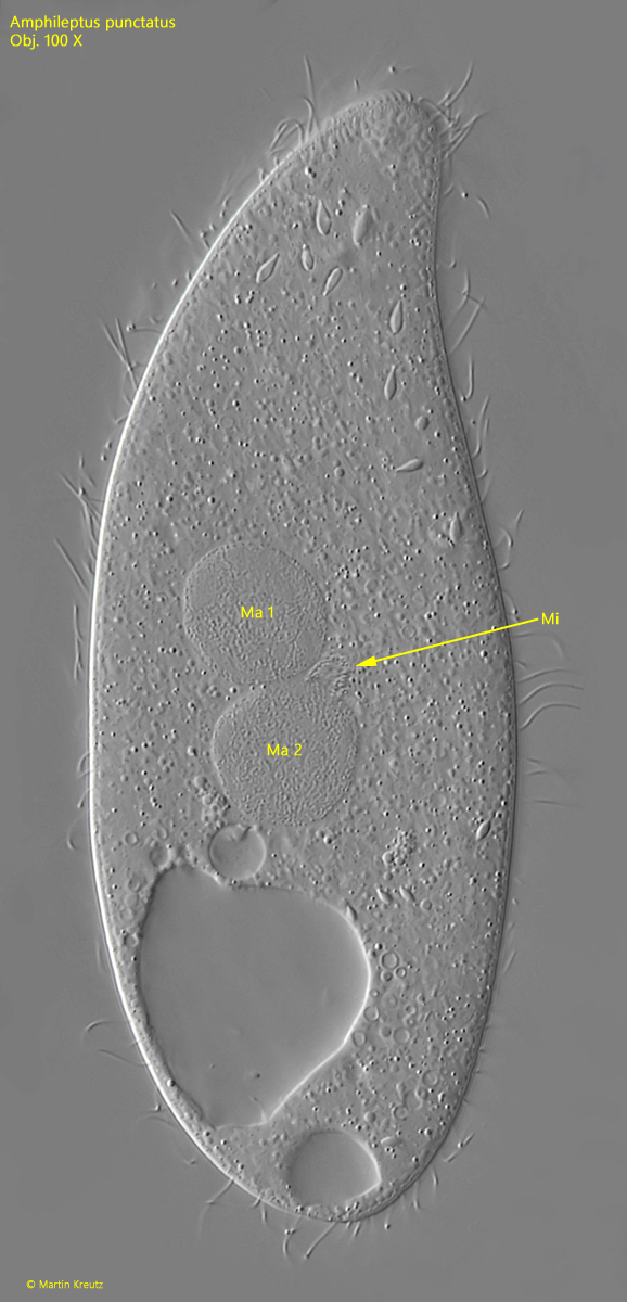
The macronucleus consists of two separate parts, between which the micronucleus is located (s. fig. 4). In my population, this was often diffuse and difficult to recognize.
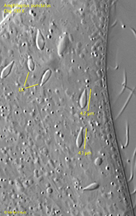
In the anterior third on the ventral side, there are usually only a few extrusomes present. At most, there are about 10, as described by Foissner et al. (1994). Often there were fewer. In my specimens, the extrusomes were about 4 µm long (s. fig. 6). These distinctively shaped extrusomes are specialized for stunning peritrich ciliates like Carchesium or Vorticella, on which Amphileptus punctatus has specialized.
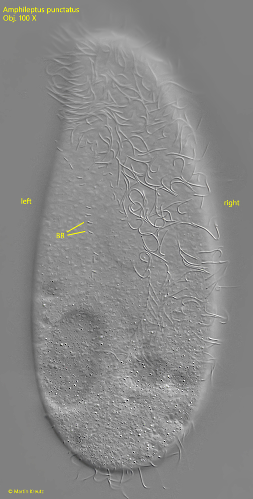
The ciliation of Amphileptus punctatus is typical for the genus. The right side is ciliated, with 18–25 rows of cilia, while the left side is bare except for 4–6 rows of short, bristle-like cilia. On the dorsal side in the anterior third, there is also a conspicuous dorsal brush visible (s. fig. 3 a-b) .
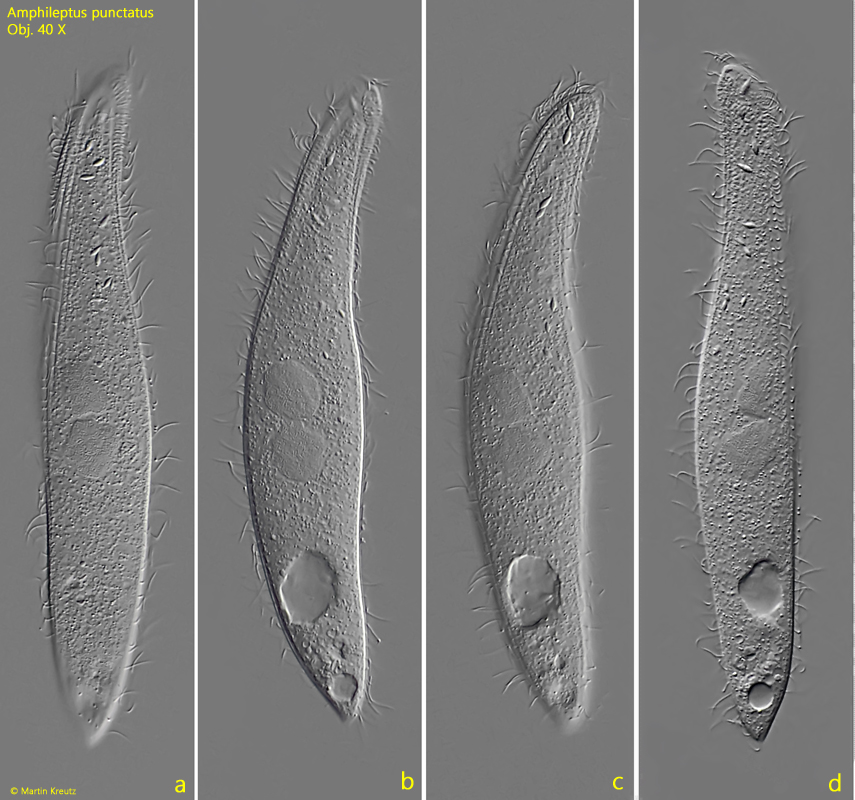
Fig. 1 a-d:Amphileptus punctatus. L = 173 µm. A freely swimming specimen from left (a-c) and from right (d). Obj. 40 X.
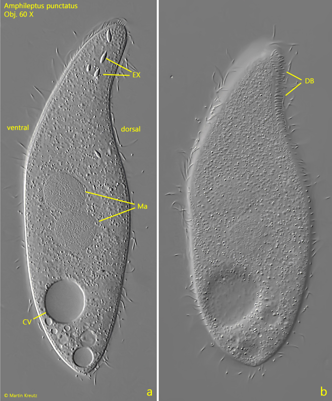
Fig. 2 a-b:Amphileptus punctatus. L = 136 µm. Two focal planes of a slightly squashed specimen from left. CV = contractile vacuole, DB = dorsal brush, EX = extrusomes, Ma = macronuclei. Obj. 60 X.
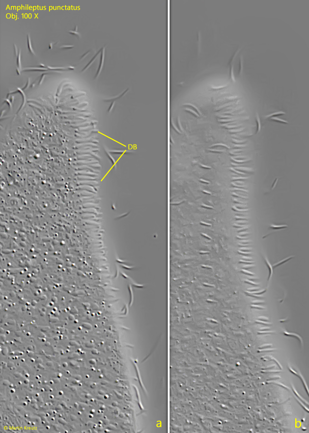
Fig. 3 a-b:Amphileptus punctatus. Two focal planes of the dorsal brush (DB). Obj. 100 X.
Fig. 4:Amphileptus punctatus. A squashed specimen from left. The micronucleus (Mi) is located between the two macronuclei (Ma 1 , Ma 2). Obj. 100 X.
Fig. 5:Amphileptus punctatus. On the left side only few rows of short, bristle-like cilia (BR) are present, while the right side is ciliated. Obj. 100 X.
Fig. 6:Amphileptus punctatus. A crop of fig. 4 with the drop-shaped extrusomes (EX) in detail. The extrusomes of this specimen have a length of about 4 µm. Obj. 100 X.
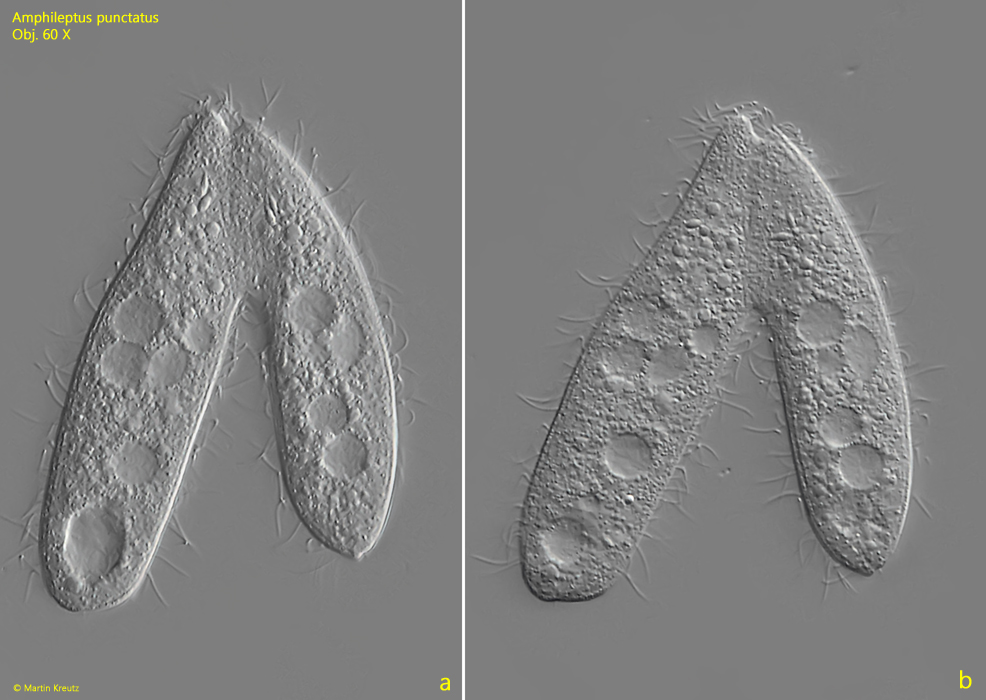
Fig. 7 a-b:Amphileptus punctatus. L = 74–85 µm. Two specimens in conjugation. Obj. 60 X.