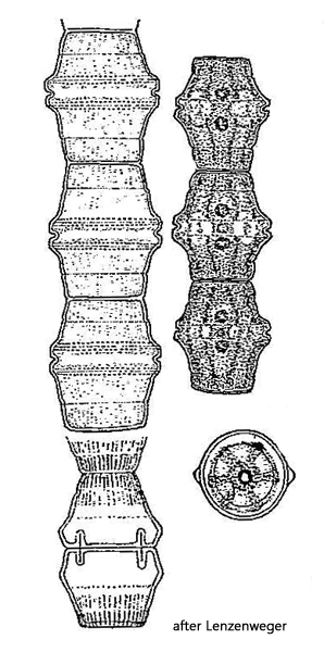
cell wall with fine longitudinal striation (hard to see)
two lobed chloroplasts with each one pyrenoid
nucleus central in a bridge of cytoplasm
Bambusina borreri
Bambusina borreri is a typical alga found in moor waters. Therefore, I have only found it so far in Austria in the Schwemm Moor and the Sima Moor. This alga is not present in my local collection sites.
The filaments of Bambusina borreri are easily recognizable by the almost hexagonal shape of the individual cells. Like almost all desmid algae, the cells consist of two mirror-symmetrical semi-cells separated by a sinus. In Bambusina borreri, however, the sinus is very rudimentary and can only be seen as a shallow groove. On both sides of the sinus, the cell wall is thickened in a bulging manner. On both sides of the sinus, there are ring-shaped rows of granules, and towards the apices, the cell wall has fine longitudinal stripes. However, this ornamentation of the cell wall is difficult to see in living specimens. Due to the typical shape of the cells, Bambusina borreri cannot be confused with any other species.
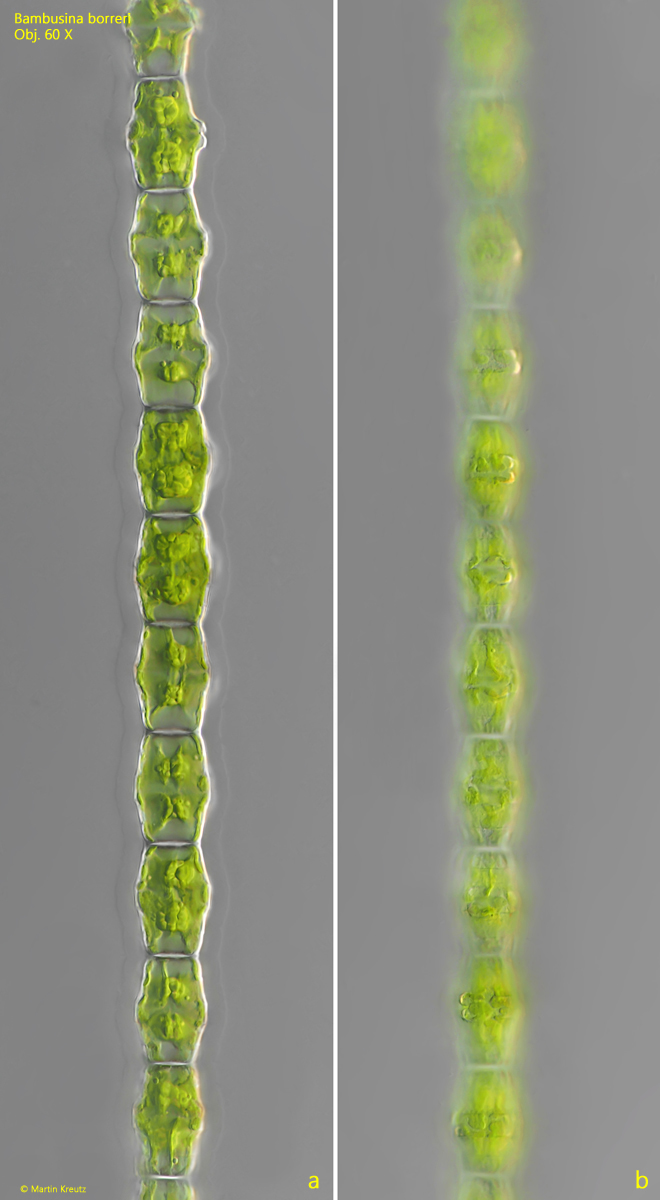
Fig. 1 a-b:Bambusina borreri. L (of cells) = 26–32 µm. Two focal planes of a filament of cells. Obj. 60 X.
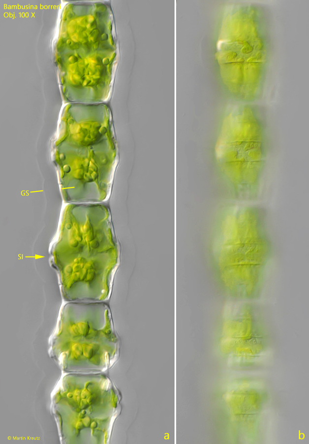
Fig. 2 a-b:Bambusina borreri. A section of the filament shown in fig. 1 a-b. The cells are covered by a gelatinous sheath (GS) with a thickness of about 10 µm. The Sinus (SI) beween the semi-cells is a weak indentation. Obj. 100 X.
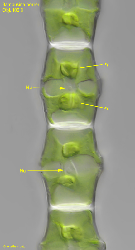
Fig. 3:Bambusina borreri. In young, transparent cells the nucleus (Nu) is visible, located in a bridge of cytoplasm. In each cell two pyrenoids (PY) are present. Obj. 100 X.
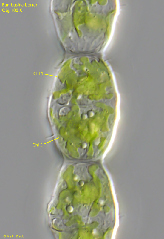
Fig. 4:Bambusina borreri. A squashed cell with focal plane on the lobed chloroplasts (Chl 1, Chl 2). Obj. 100 X.