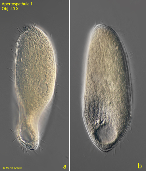
In July 2005, I found a brownish-colored ciliate in the top layer of mud in the Simmelried, which was about 140 µm long, swam slowly, and rotated around its longitudinal axis. The shape was compact spindle-shaped with a terminal contractile vacuole. While swimming, an oral bulge could be faintly seen. In the following years, I found a few more specimens in August 2008, January 2022, May 2022, and July 2024. All specimens came from the Simmelried.
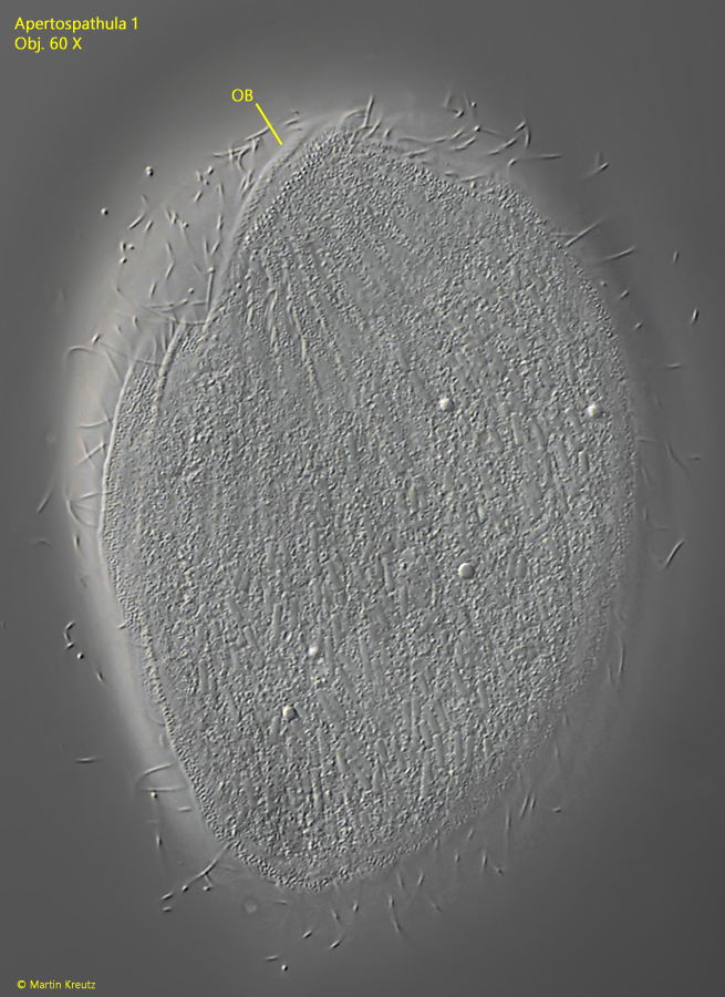
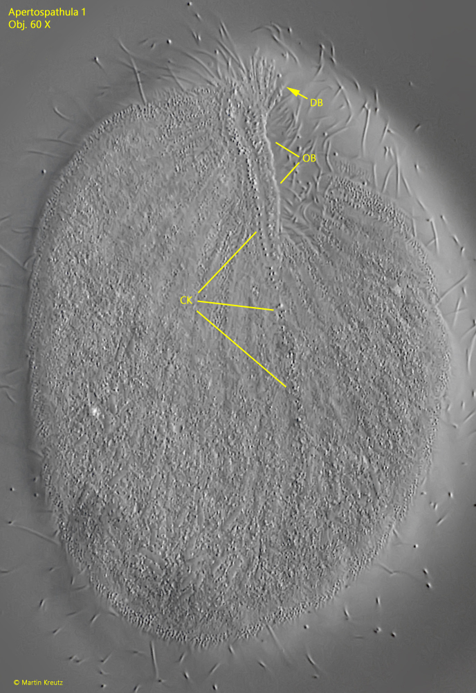
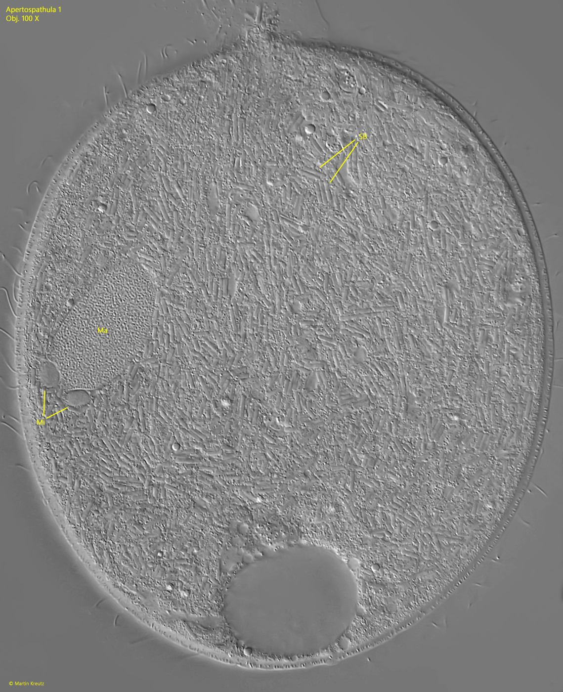
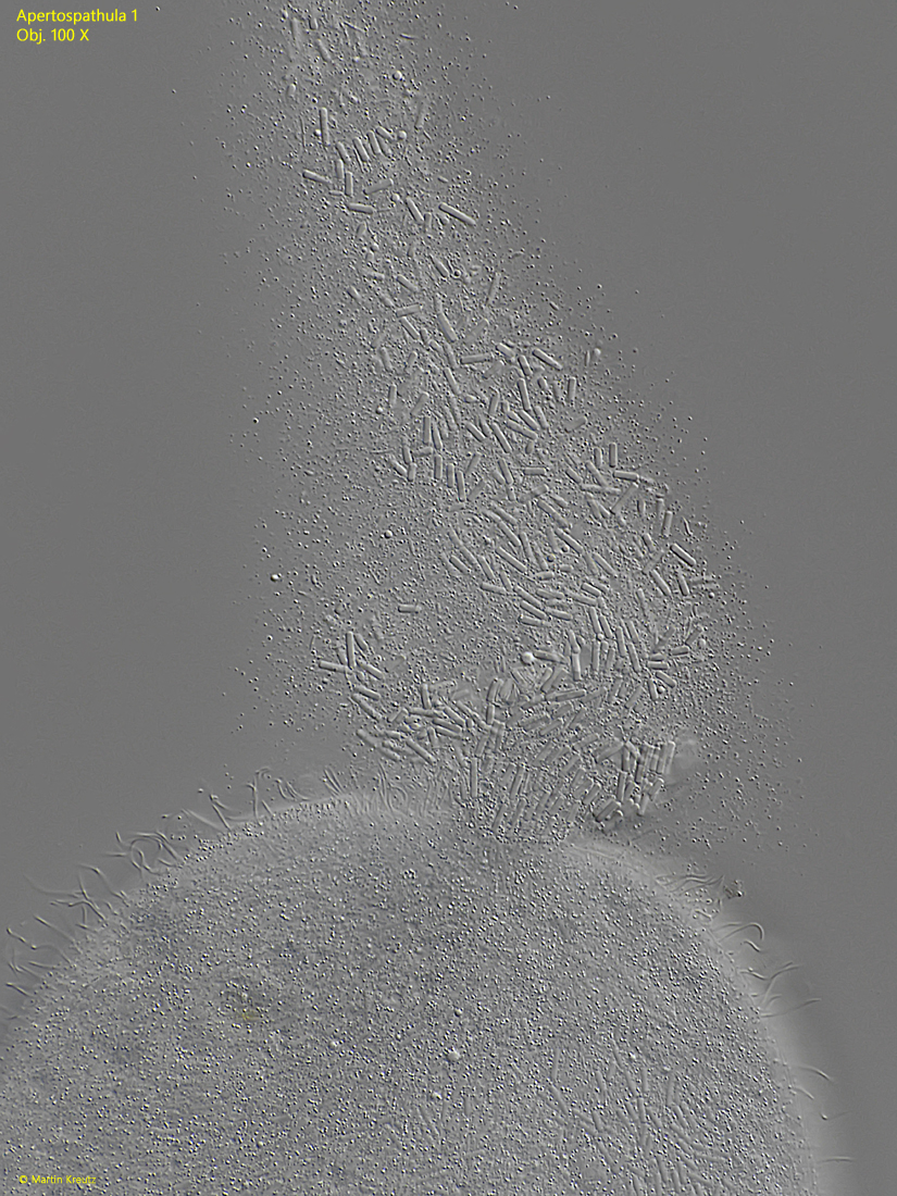
Closer examination of the specimens revealed that it is a spathidiid ciliate of the genus Apertospathula, which has not yet been given a taxonomic species name. The genus Apertospathula is characterized by a circumoral kinety of cilia that is not closed ventrally. Instead, it is extended ventrally on the right side opposite the left side (s. fig. 4).
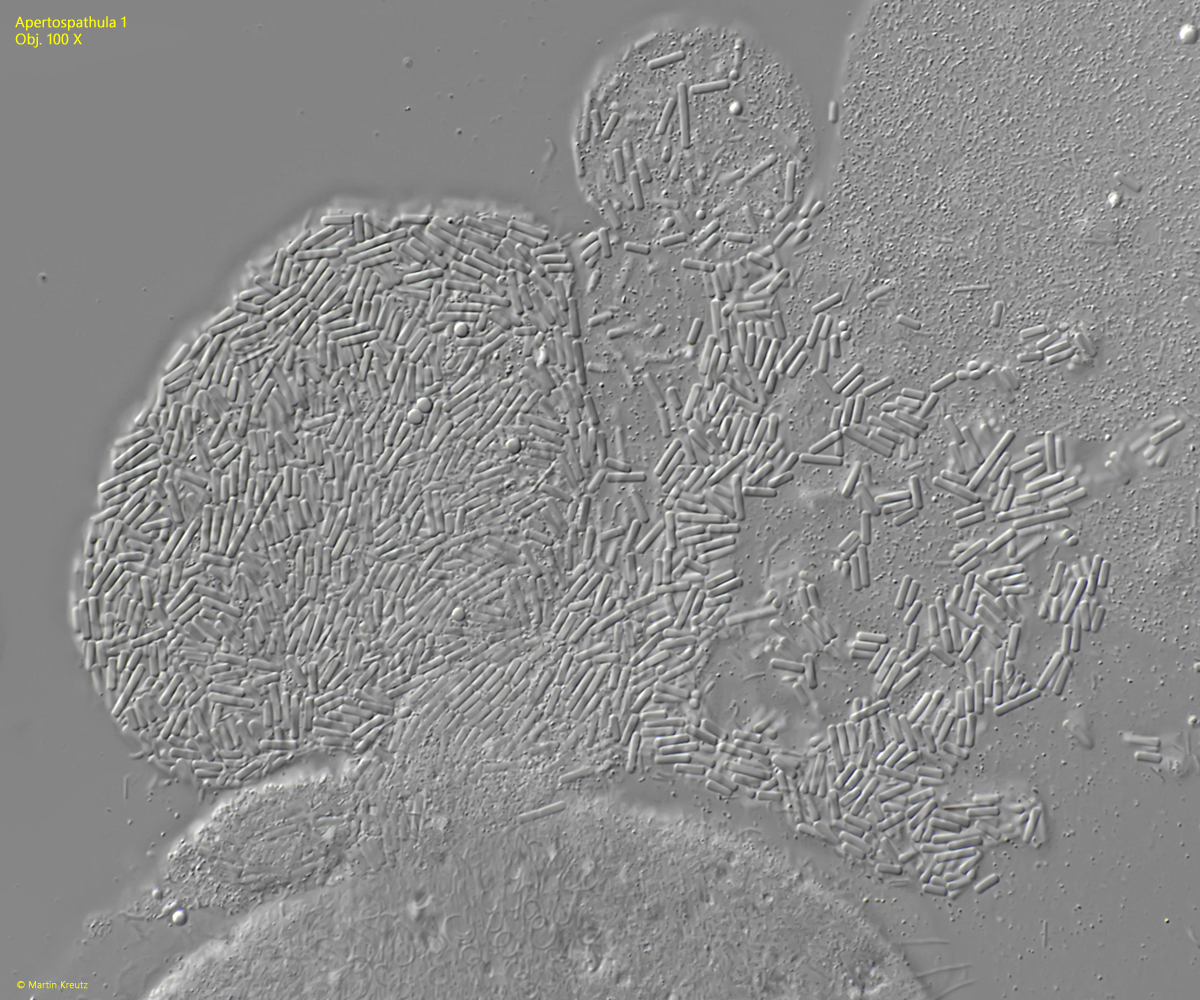
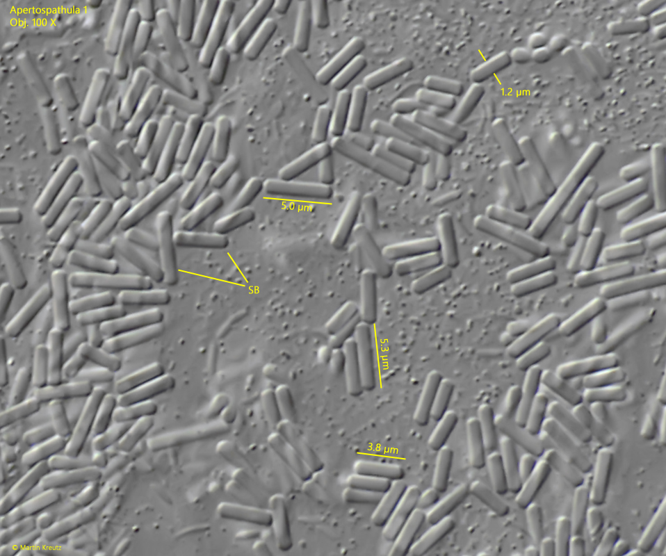
The reason for the opaque cytoplasm of this ciliate only becomes apparent when the specimens are strongly compressed. Then it can be seen that the entire body is densely filled with bacteria of the same species (s. fig. 5). They are distributed in the cytoplasm and not in food vacuoles. It is therefore assumed that these are endosymbionts, since all the bacteria are also of the same species. With further increasing coverslip pressure, the bacteria are released and can then be examined more closely (s. figs. 6 and 7). They are rods with a constant diameter of 1.2 µm and a length of 4–5 µm (s. fig. 8). I have not been able to observe such a high concentration of endosymbiotic bacteria in any other ciliates so far.
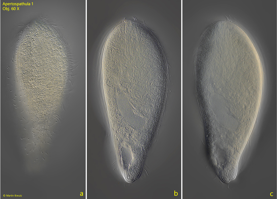
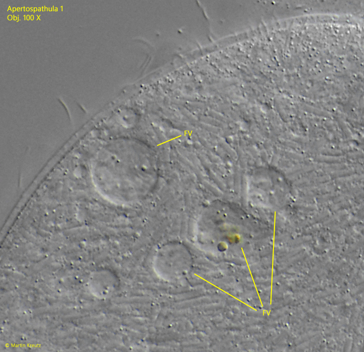
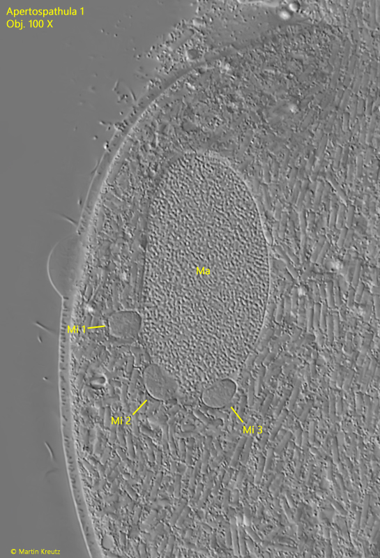
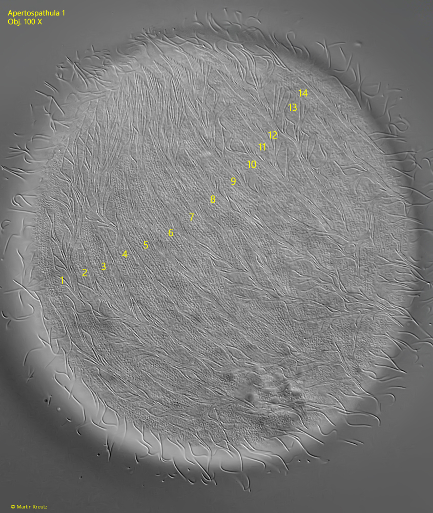
The specimens of my population had an ellipsoid or kidney-shaped macronucleus and three spherical micronuclei each, which were adjacent to the macronucleus (s. fig. 10). The length of the specimens ranged between 113–144 µm. Only very few food vacuoles with an unidentifiable content could be identified in the specimens (s. fig. 9). It cannot be ruled out that part of the symbiotic bacteria are also cyclically subjected to digestion in order to maintain the population constant. In compressed specimens, I was able to identify 14–15 somatic kineties on one side of the body, so that 28–30 are present around the entire body (s. fig. 11).
This ciliate was also found by other authors:
The documentation and descriptions of these authors differ from my observations in only a few points. Weiss & Esteban report the length of the symbiotic bacteria as only 2–3 µm and the number of somatic kineties in their population as 10–16. Additionally, their specimens were larger, measuring 150–200 µm. In Qassis’ population, the macronucleus was almost U-shaped with only one micronucleus. Despite these differences, it can be assumed that the specimens examined by Qassis and Weiss & Esteban belong to the same species Apertospathula as in my population due to the high concentration of symbiotic bacteria in the cytoplasm, which makes this ciliate unmistakable.