akinetes formed by severeal cells in common, brown cell wall
cells blueish-green with granules in cytoplasm
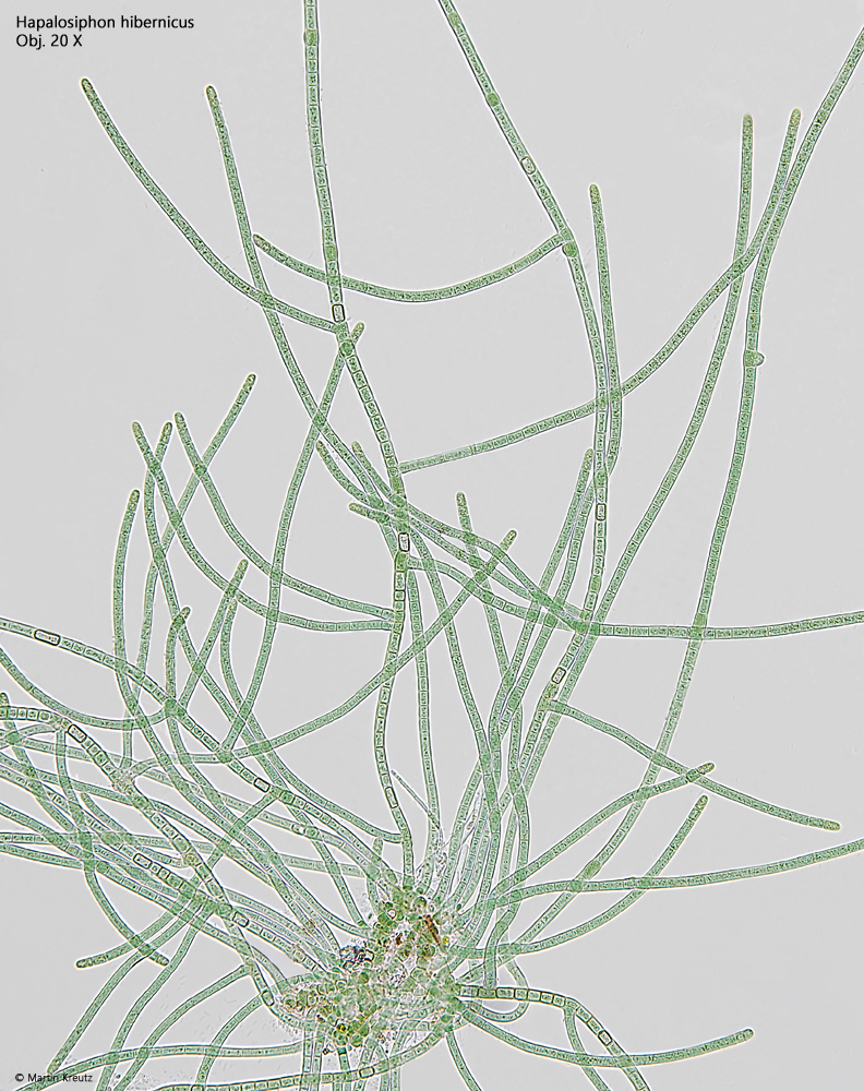
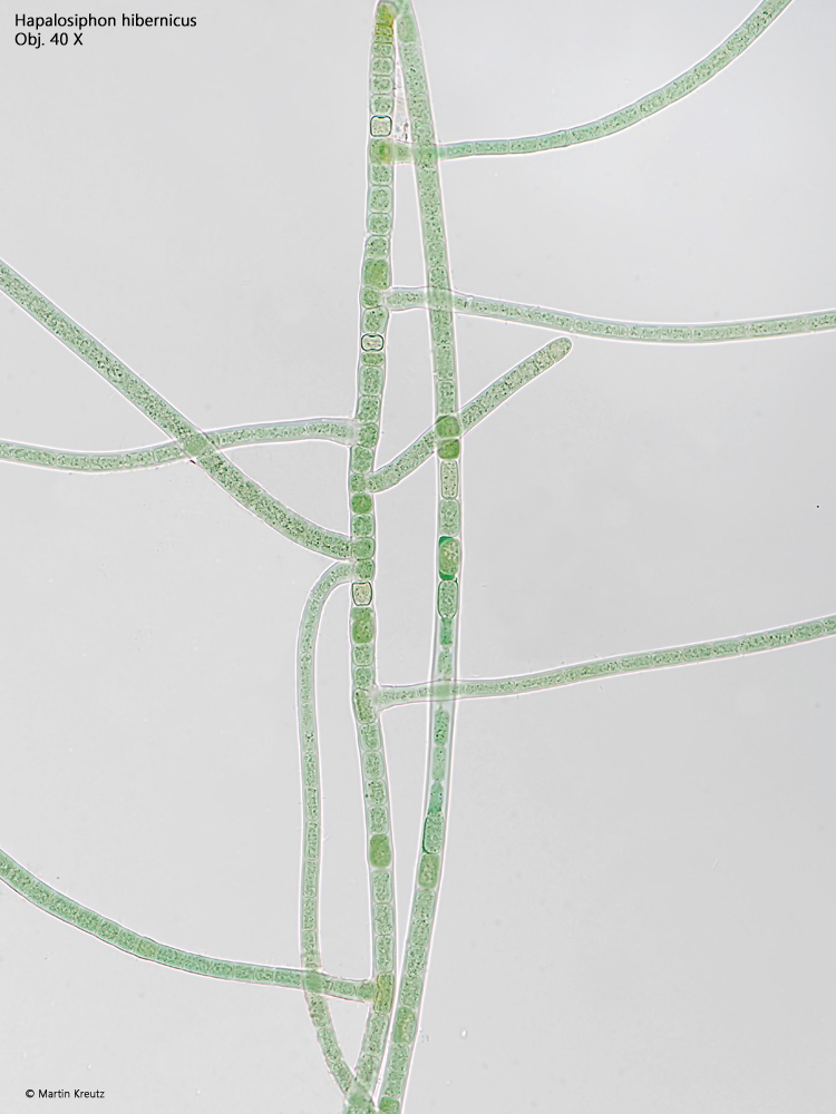
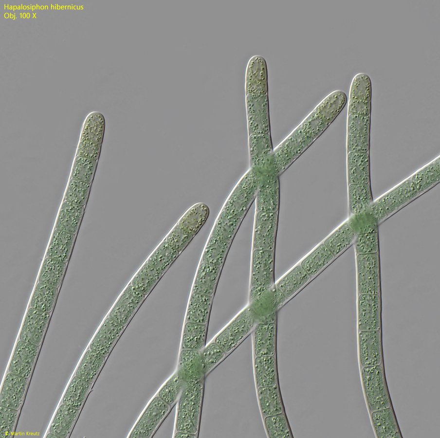
Hapalosiphon hibernicus
So far, I have only found Hapalosiphon hibernicus in the Lauchsee Moor in Austria. The branched filaments grew in large numbers on the vessel wall after a few weeks.
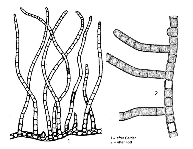
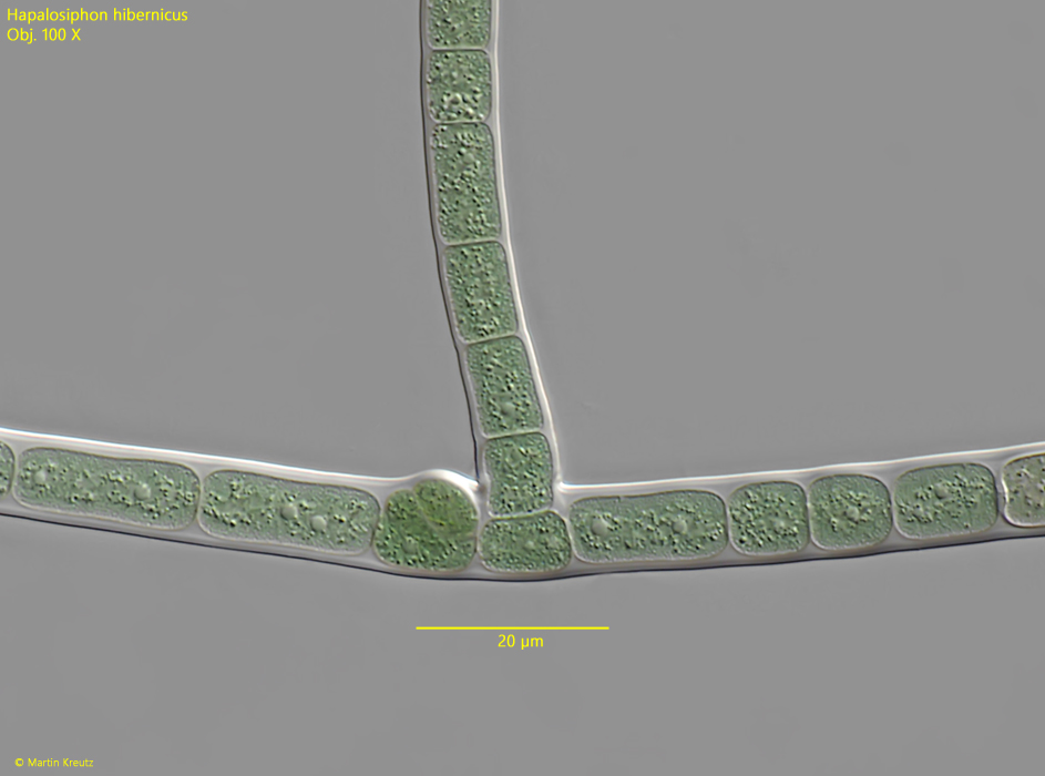
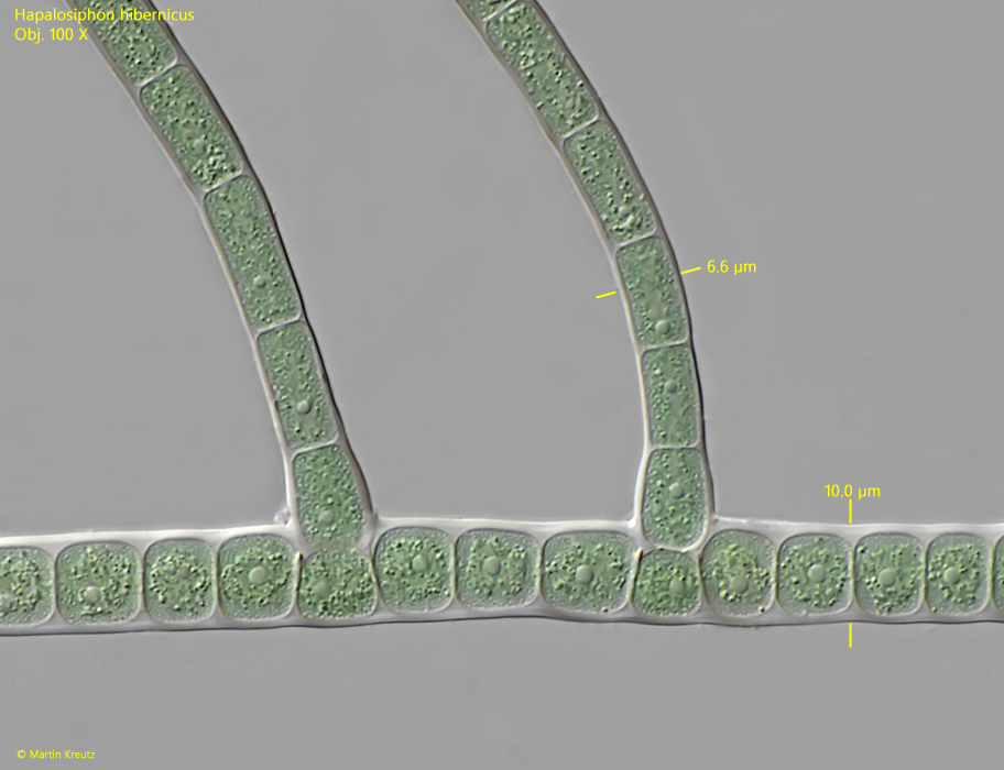
Hapalosiphon hibernicus can be relatively well recognized by its typical growth form, as the branching filaments extend at a 90° angle from the main filament (s. figs. 1 and 2). These are true branches because after a cell in the filament divides parallel to the growth direction of the filament, one of the parallel daughter cells continues to divide and then forms the branching filament. The second daughter cell remains in the main filament. This arrangement can be clearly seen at every branching point (s. figs. 4 and 5).
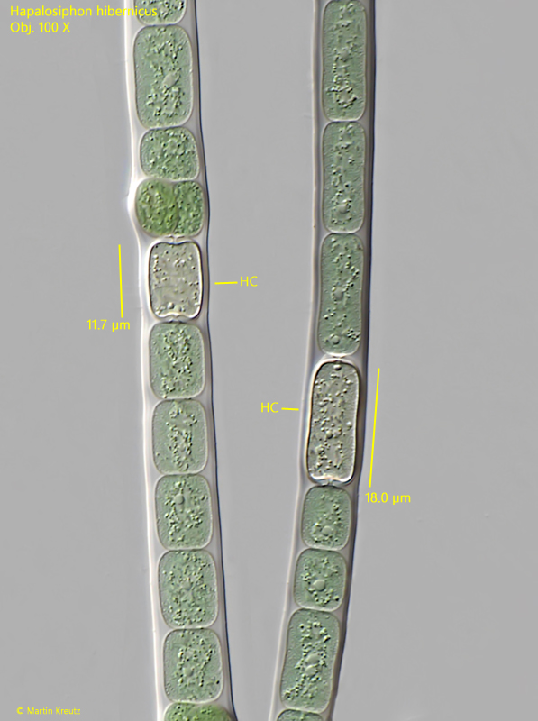
The vegetative cells are all rectangular and usually between 5–6.5 µm wide. However, they do not have a constant length within the filament. Scattered among the vegetative cells in the filaments are also rectangular heterocysts. These have a thickened cell wall and are somewhat lighter in color than the blue-green vegetative cells (s. fig. 6). I could not observe the formation of akinetes from several cells within a thickened, common cell wall.
Fig. 1:Hapalosiphon hibernicus. L = about 650 µm (of filaments). Overview of some branched filaments. Obj. 20 X.
Fig. 2:Hapalosiphon hibernicus. Detail of the branched filaments in brightfield illumination. Obj. 40 X.
Fig. 3:Hapalosiphon hibernicus. D = 6.2–6.5 µm (of filaments). The terminal cells of the filaments are slightly tapered and yellowish. Obj. 100 X.
Fig. 4:Hapalosiphon hibernicus. A branching in the detail. The main filament has a diameter of 10 µm while the branches have 6.6 µm. Obj. 100 X.
Fig. 5:Hapalosiphon hibernicus. A second filament with the branches in detail. Obj. 100 X.
Fig. 6:Hapalosiphon hibernicus. The rectangular heterocysts (HC) with a thickened cell wall are scattered in the filamants between the vegetative cells. Obj. 100 X.