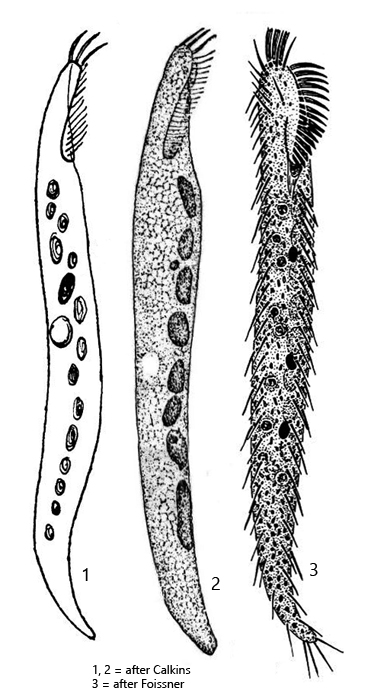
body slender vermiform, apically slightly flattened
length 94–283 µm, width 12–35 µm
widely spaced cirri
adoral zone only 10–15 % of body length
3 frontal cirri
6–26 macronuclear nodules, nodules left of midline
about 17 longitudinal rows of yellowish or colorless cortical granules
contractile vacuole near mid-body, left side
transverse and caudal cirri absent
Engelmaniella mobilis
So far, I have found Engelmaniella mobilis exclusively in the Simmelried, where the ciliate is very rare. I have only found it twice, in November 2016 and November 2020. In total, however, there were only 3 specimens.
At low magnification, the slender, vermicular body of Engelmaniella mobilis resembles some slender representatives of the genus Chaenea. At medium magnification, however, the adoral zone is clearly visible, which accounts for only 10-15% of the body length (s. fig. 2 a). For a hypotrich ciliate, the ciliation is greatly reduced. Widely spaced cirri are distributed over the body, but they are thin and soft. Transverse and caudal cirri are completely absent. Only at the front end are three distinct frontal cirri present (s. fig. 3 d). The macronucleus consists of several nodules that lie along the longitudinal axis in the body. The contractile vacuole is located in the middle of the body.
The few specimens of my population were between 105–256 µm long. The body length of Engelmaniella mobilis seems to be very variable, ranging from 94–283 µm, and has been measured by various authors in 7 different populations. The average across all populations is 125 µm body length. The nodules of the macronucleus were difficult to see in all specimens of my population because the specimens were densely filled with highly refractive granules (s. figs. 1 b and 4). The contractile vacuole was located in the middle of the body in all specimens (s. figs. 1 d and 2 d). I could not recognize any rows of midventral cirri on the ventral side, as would be typical for the related genus Uroleptus. I could not detect any rows of cortical granules on my specimens, as mentioned by Berger (2011). Otherwise, all features corresponded to those of Engelmaniella mobilis.
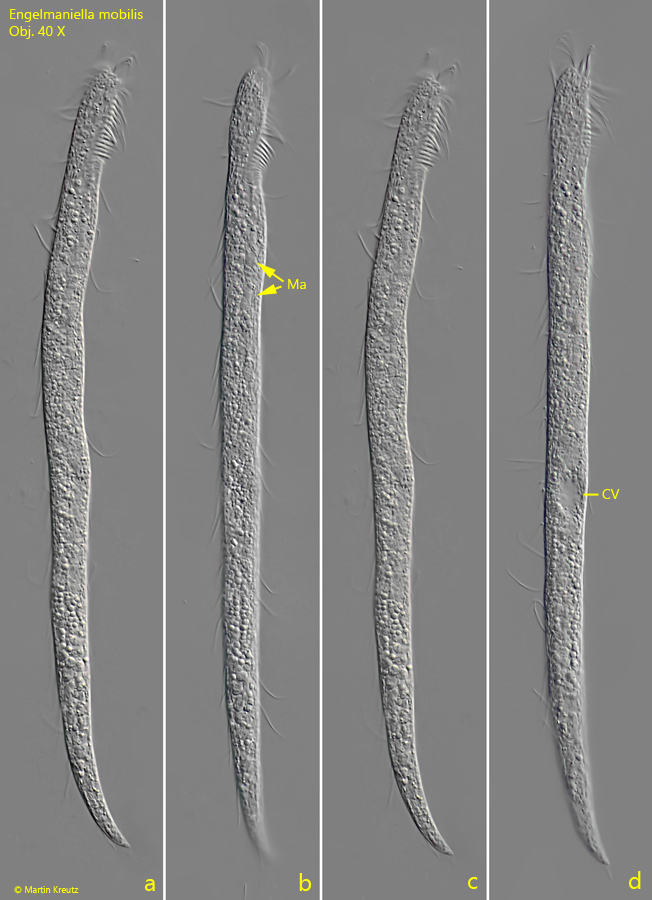
Fig. 1 a-d:Engelmaniella mobilis. L = 256 µm. A freely swimming specimen from ventral (a, b) and from right (c, d). The nodules of the macronucleus (Ma) are hard to see between the refractive granules filling the cytoplasm. CV = contractile vacuole. Obj. 40 X.
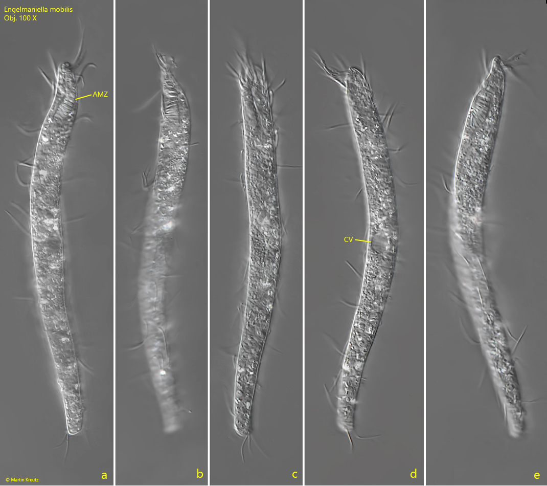
Fig. 2 a-e:Engelmaniella mobilis. L = 128 µm. A second, freely swimming specimen found in November 2016 in the Simmelried. The body is covered with widely spaced cirri. The anterior end is slightly flattened and gives a snout-like impression (e). CV = contractile vacuole. Obj. 100 X.
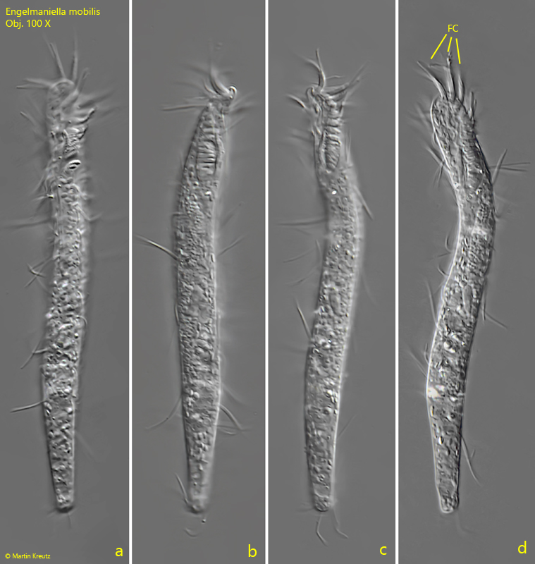
Fig. 3 a-d:Engelmaniella mobilis. L = 105 µm. A third specimen found in December 2016 in the Simmelried. Note the three distinct frontal cirri (FC). Obj. 100 X.
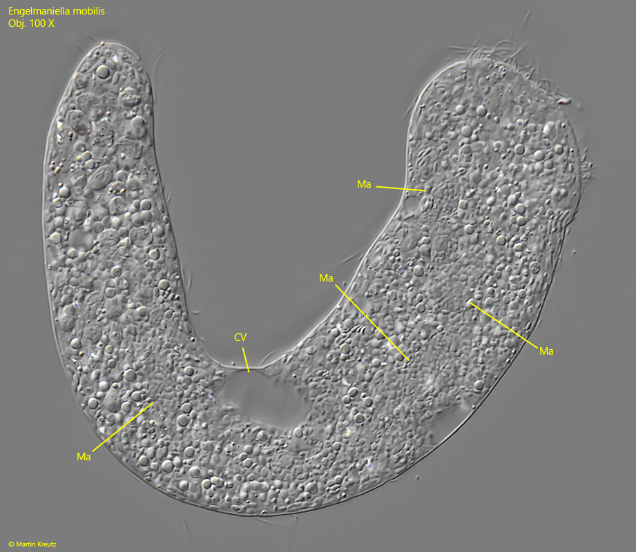
Fig. 4:Engelmaniella mobilis. The squashed specimen as shown in fig. 1 a-d. The nodules of the macronucleus (Ma) are hard to see between the granules filling the cytoplasm. CV = contractile vacuole. Obj. 100 X.