Synonyms:Encyonema cespitosum var. auerswaldii, Cymbella ventricosa var. auerswaldii, Cymbella cespitosa var. auerswaldii, Cymbella prostrata var. auerswaldii, Cocconema cespitosum var. auerswaldii
I found Encyonema auerswaldii in large quantities on the stems of the yellow water lily (Nuphar lutea) in the pond of the waste disposal company Constance. The tube-shaped colonies were already macroscopically visible as 1–2 mm long, dark brown filaments on the stems. They could be easily removed by scraping them off the stems and were then easy to examine.
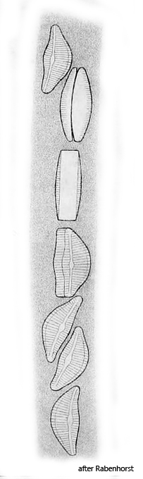
Encyonema auerswaldii forms a tube-shaped, gelatinous tube with a diameter of about 30–40 µm in which cell division takes place. This causes the tube to grow continuously and it can also branch. The cells are approximately semi-circular in valve view and approximately rectangular in girdle view. The raphe on the valve side is straight and only bends at the apices to the dorsal, convex side. In addition, the raphe is shifted towards the slightly convex, ventral side. The nucleus is centrally located and has a distinct, central nucleolus.
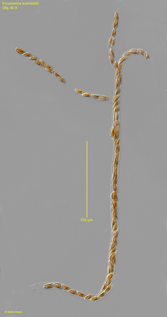
Fig. 1:Encyonema auerswaldii. L = about 1 mm (of colony). A branched colony of about 100 cells in a gelatinous tube. Obj. 40 X.
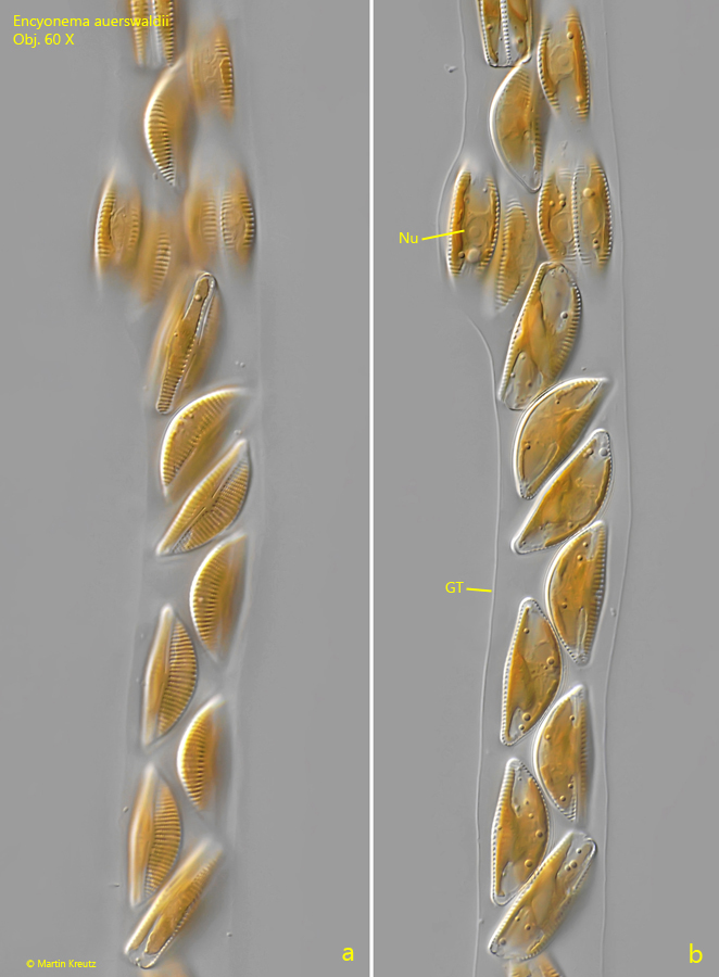
Fig. 2:Encyonema auerswaldii. L = 36–38 µm (of cells). A section of the colony as shown in fig. 1. Obj. 60 X.
Fig. 3:Encyonema auerswaldii. L = 36–39 µm (of cells). A second section of the colony as shown in fig. 1. Note the nucleus (Nu) of the cells with a central nucleolus. GT = gelatinous tube. Obj. 60 X.
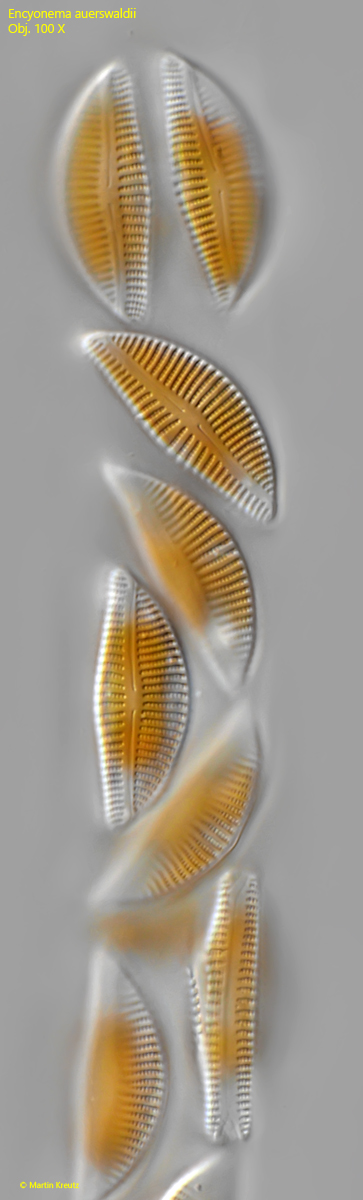
Fig. 4:Encyonema auerswaldii. L = 36–38 µm (of cells). Some cells in the gelatinous tube in valve view. Obj. 100 X.
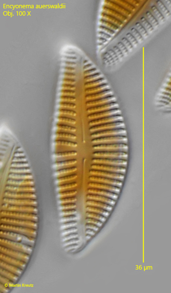
Fig. 5:Encyonema auerswaldii. L = 36 µm. The valve view of a cell in detail. Obj. 100 X.