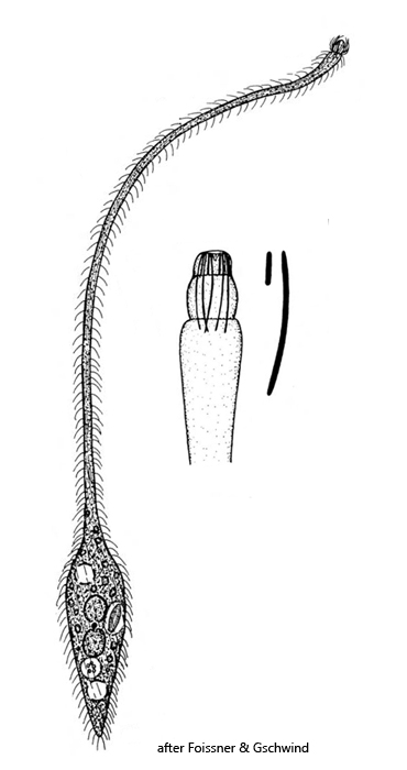
neck thin, stretchable to several times of body length
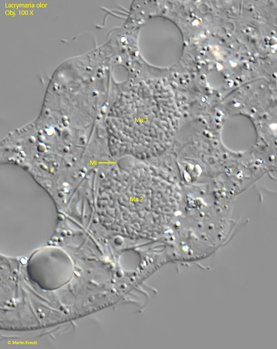
two ellipsoid macronuclei, one micronucleus in between
one subterminal contractile vacuole, a second one near anterior end
13–16 spirally rows of cilia, running counterclockwise
head conically shaped with spirally rows of long cilia
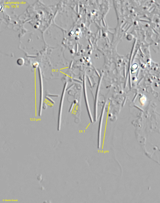
head and cytoplasm filled with 2–3 µm and 10–12 µm long extrusomes
Lacrymaria olor
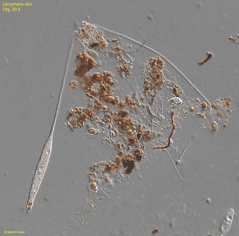
I do find Lacrymaria olor at many of my sampling sites, but mostly only single specimens. In my experience, the species seems to be more often found among dead aquatic plants floating on the surface.
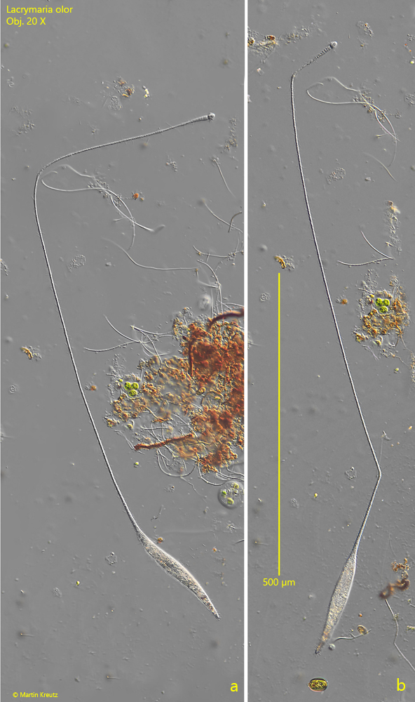
The most characteristic feature of Lacrymaria olor is the high contractility and mobility of this species. This especially concerns the neck, which can stretch up to 800 µm in length. Although the neck becomes very thin in the process, it is extremely flexible and mobile. It often bends at angles but can also bend almost circularly (s. fig. 2 a-b). The head, armed with extrusomes, performs very fast, searching, and tactile movements.
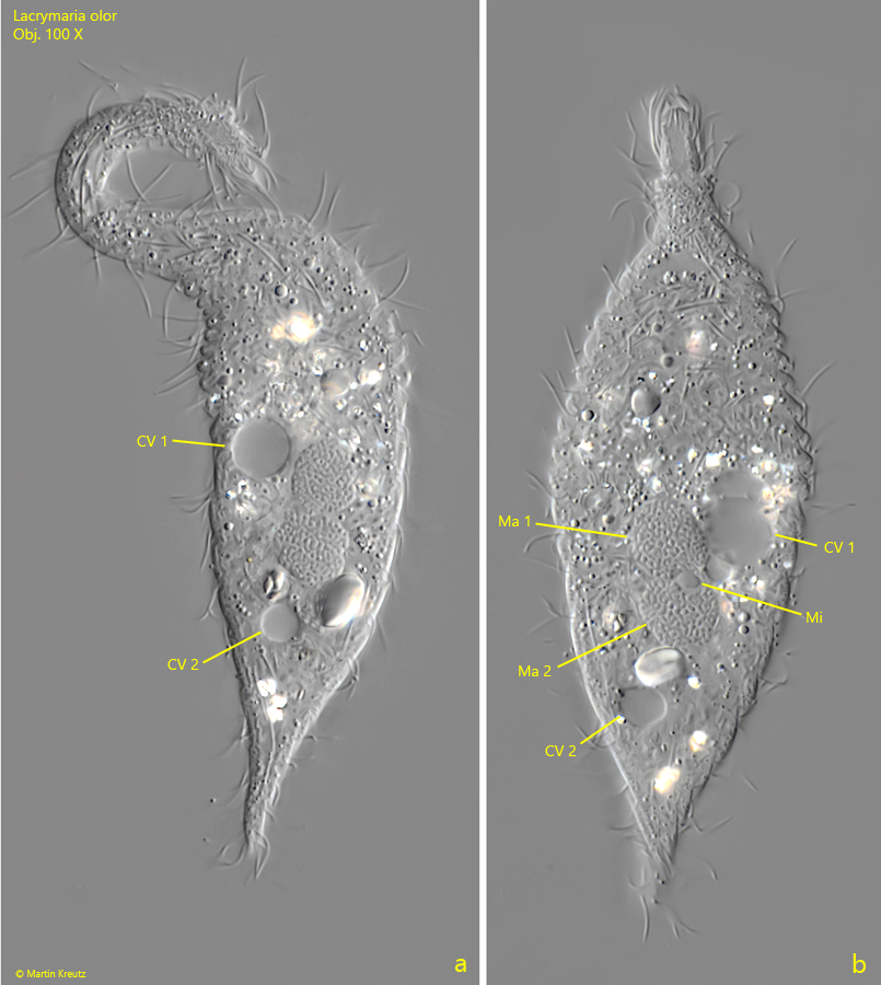
Lacrymaria olor differs from the numerous other species within the genus Lacrymaria by the two macronuclei and two contractile vacuoles. The two macronuclei are located approximately in the middle of the body. Between them lies a small micronucleus (s. figs. 5 b and 8). Of the two contractile vacuoles, one is subterminal at the posterior end while the second is found in the anterior third of the body (s. fig. 5 a).
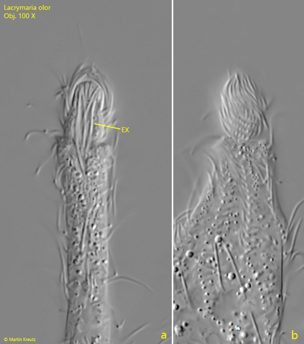
The longitudinal rows of cilia run counterclockwise over the body (s. fig. 6). Between the rows of cilia are rows of granules. Spiral rows of elongated cilia are also present on the head (s. fig. 7 b). These form an apical tuft of cilia that protrudes beyond the apical end of the head. In the head of Lacrymaria olor, there are 2 types of extrusomes, which are also distributed in the cytoplasm. Type 1 extrusomes are slightly curved rods with a length of 11–12 µm, and type 2 are very short, very slightly curved rods with a length of 2–3 µm (s. figs. 7 a and 9).
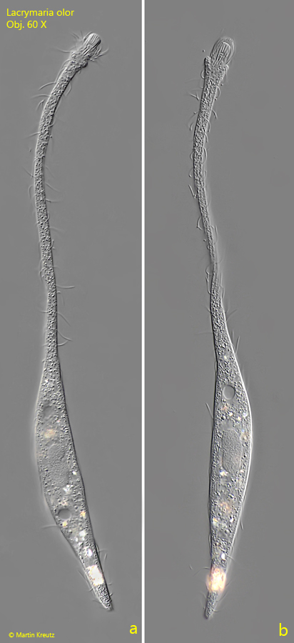
Fig. 1 a-b:Lacrymaria olor. L = 210 µm. A slighly elongated specimen. Obj. 60 X.
Fig. 2 a-b:Lacrymaria olor. L = ~ 900 µm. A fully elongated specimen. Obj. 20 X.
Fig. 3:Lacrymaria olor. L = ~700 µm. An elongated specimen searching for prey behind an obsticle. Obj. 20 X.
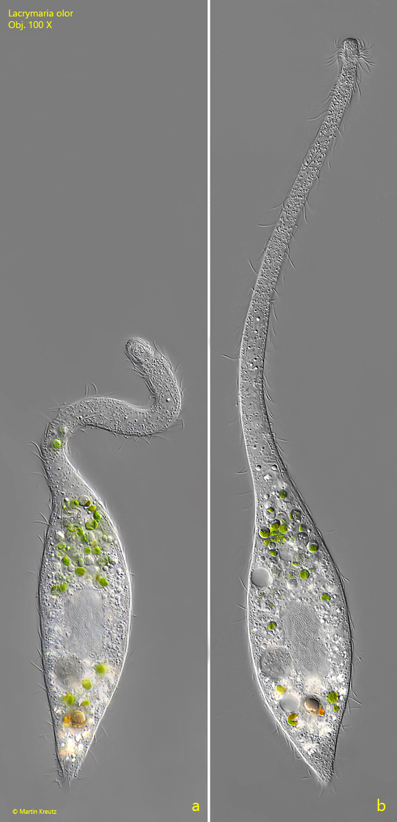
Fig. 4 a-b:Lacrymaria olor. L = 180 µm. A slightly squashed specimen. Obj. 100 X.
Fig. 5 a-b:Lacrymaria olor. L = 120 µm. A slightly squashed specimen. The two macronuclei (Ma 1, Ma 2) with the micronucleus (Mi) in between is visible and the two contractile vacuoles (CV 1, CV 2). Obj. 100 X.
Fig. 6:Lacrymaria olor. L = 130 µm. Focal plane on the spirally rows of cilia running counterclockwise along the body. Obj. 100 X.
Fig. 7 a-b:Lacrymaria olor. Two focal planes of the conical head filled with extrusomes (EX). The rows of elongated cilia of the head are running also counterclockwise (b). Obj. 100 X.
Fig. 8:Lacrymaria olor. The two macronuclei (Ma 1, Ma 2) and the micronucleus (Mi) in a squashed specimen. Obj. 100 X.
Fig. 9:Lacrymaria olor. In a strongly squashed the 2 types of extrusomes are visible. The larger type 1 extrusomes (EX 1) are slightly curved and 11–12 µm long while the smaller type 2 extrusomes (EX 2) are also slightly curved with a length 0f 2–3 µm. Obj. 100 X.