Radiofilum mesomorphum Skuja, 1956

Most likely ID: n.a.


Synonym: n.a.


Sampling location: Simmelried, Lauchsee Moor (Austria)


Phylogenetic tree: Radiofilum mesomorphum


Diagnosis: 

  • cells globose or rounded-lentiform
  • cells are arranged in single series in an unbranched filament
  • filament in a gelatinous sheath, 33–40 µm thick
  • length 5–14 µm, width 13–17 µm (of cells)
  • cells wall of 2 overlapping pieces, equatorial thickening
  • one chloroplast, parietal
  • one pyrenoid
  • starch granules scattered in cytoplasm
  • cell divisions parallel to longutinal axis of filament
Radiofilum-mesomorphum
Radiofilum mesomorphum

So far, I have found Radiofilum mesomorphum only a few times in the Simmelried and in a sample from the Lauchsee Moor in Austria. In my other locations, I have not been able to detect this alga so far.


Radiofilum mesomorphum is essentially recognized by the somewhat lens-shaped cells, which line up in a row to form a filament and are surrounded by a thick, gelatinous mucilage sheath, which changes its thickness depending on the position of the cells in the filament. At high magnification, a thickening can be seen at the equatorial margins of the cells. These are caused by the structure of the cell wall, which consists of two cup-shaped parts that also overlap slightly. This structure is best observed during cell division, when these two shells separate and the daughter cells regenerate the missing halves (s. fig. 4).

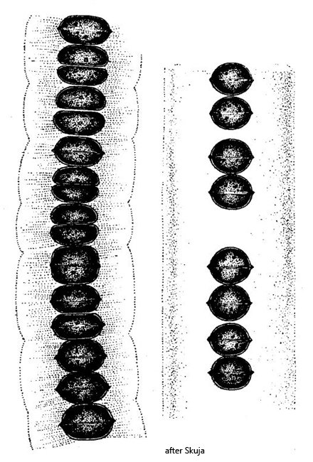
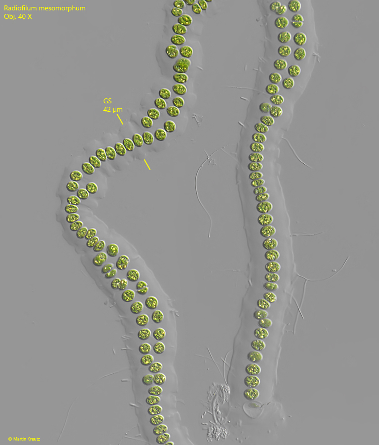
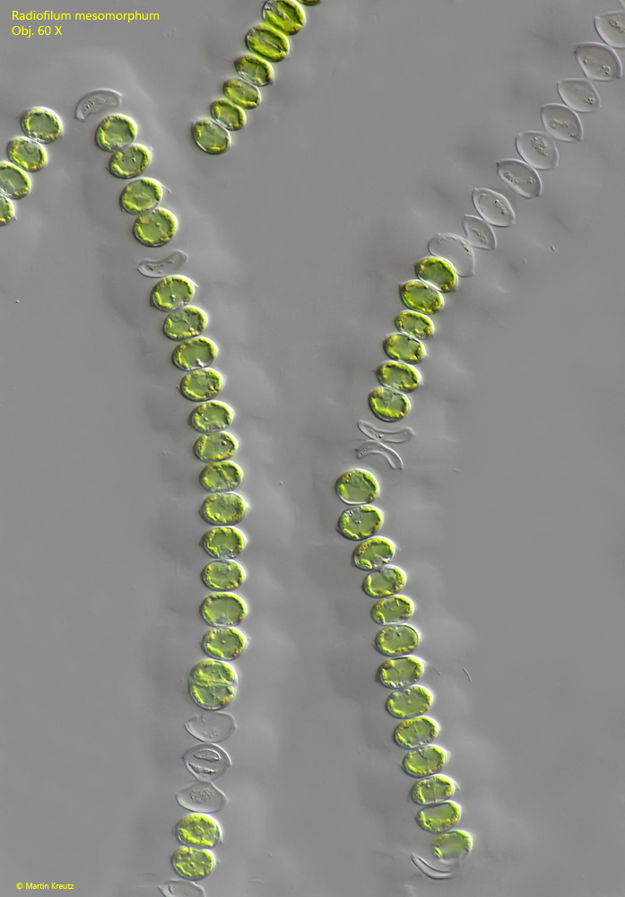
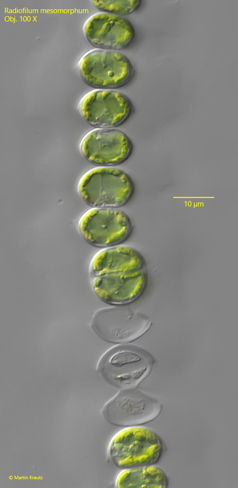
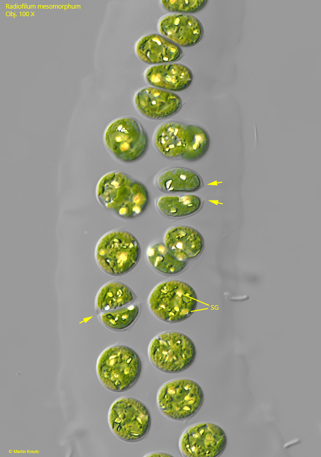
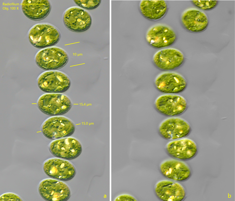
The cells of my population were between 12–15.5 µm wide and about 7–11 µm long. Only a few cells were nearly spherical. Based on the size of the cells, this must be Radiofilum mesomorphum. This species was established by Skuja (1956) after he determined that the cells of Radiofilum mesomorphum are about twice as large as those of the similar species Radiofilum conjunctivum. Their cells only reach a width of about 6 µm. Therefore, Skuja separated the larger form as its own species, Radiofilum mesomorphum.
Radiofilum-mesomorphum

Fig. 1: Radiofilum mesomorphum. Two filaments found in September 2025 in the Lauchsee Moor. The gelatinous sheath (GS) of the filament is about 40 µm thick. Obj. 40 X.

Radiofilum-mesomorphum

Fig. 2: Radiofilum mesomorphum. Two filaments found in April 2022 in the Simmelried. Obj. 60 X.

Radiofilum-mesomorphum

Fig. 3: Radiofilum mesomorphum. A section of a filament as shown in fig. 2 in detail. The width of the cells is 12.4–13.2 µm. Obj. 100 X.

Radiofilum-mesomorphum

Fig. 4: Radiofilum mesomorphum. A section of a filament with a double row of cells. In dividing cells the margins of the two cup-shaped parts of the mother cell are visible (arrows). SG = starch grains. Obj. 100 X.

Radiofilum-mesomorphum

Fig. 5 a-b: Radiofilum mesomorphum. Two focal planes of some cells in a filemant. The cells have a width of 14.5–15.5 µm and a length of about 10 µm. Obj. 100 X.