Anuraeopsis fissa Gosse, 1851

Most likely ID: n.a.


Synonym: n.a.


Sampling location: Simmelried, Ulmisried, Pond of the waste disposal company Constance, Schwemm Moor (Austria)


Phylogenetic tree: Anuraeopsis fissa


Diagnosis: 

  • body oval or ton-shaped
  • ventral side flat, dorsally convex
  • cuticle smooth
  • length 80–120 µm
  • often colored yellowish or yellowish-brown
  • one large eyespot
  • amictic eggs large, drop-shaped, brown or yellowish brown
  • egg is carried in front of ventral side
  • posteriorly a protrusible organ (Weber organ)
Anuraeopsis-fissa
Anuraeopsis fissa
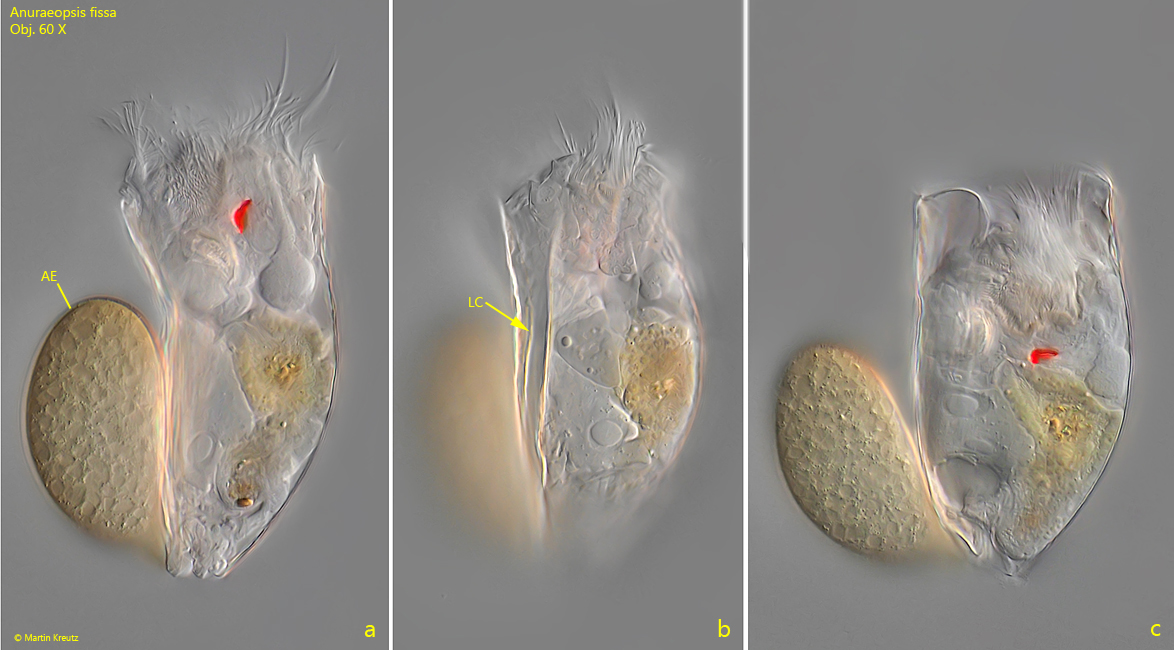
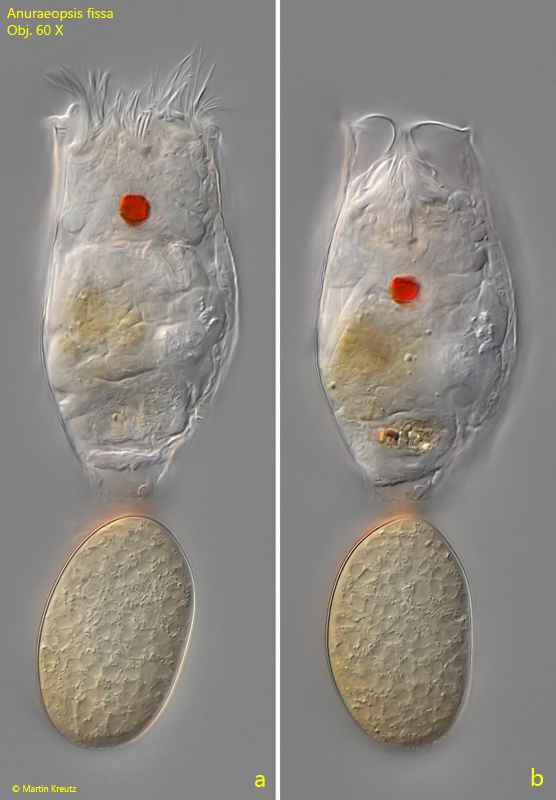
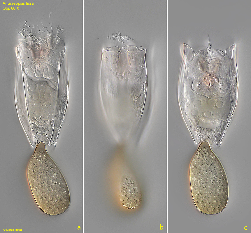
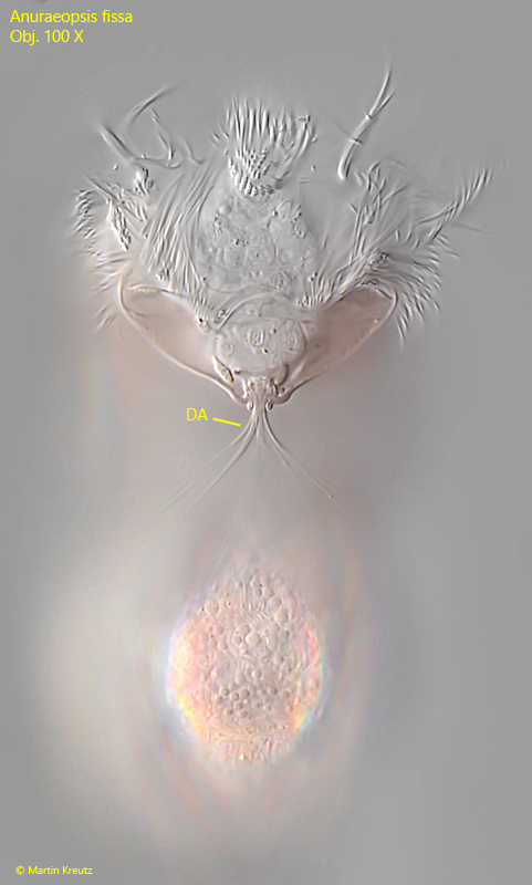
I find Anuraeopsis fissa in the plankton and among floating aquatic plants at many of my sampling sites. The species is easily recognizable by the smooth cuticle without ornamentation, which is a clear difference from the similarly shaped species Keratella cochlearis. The eyespot of Anuraeopsis fissa is very large, and specimens are often found carrying a very large, drop-shaped egg on the ventral side of the body (s. fig. 1 a). It is connected to the body by a short papilla at the posterior end. It can also be folded up and down with the help of this papilla. According to Koste (1978), the species is described as yellowish to yellowish-brown in color. The specimens of my population were very lightly pink-colored.

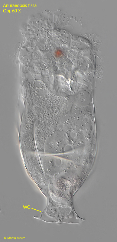
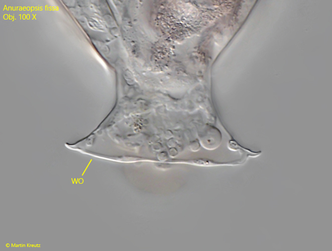
As a special feature, Anuraeopsis fissa has an extendable organ with hooks at the posterior end. I was only able to see it in squashed specimens. It is said to serve for attaching the excreted egg to the body.
 

More images and information on Anuraeopsis fissa: Michael Plewka-Freshwater life-Anuraeopsis fissa

Anuraeopsis-fissa

Fig. 1 a-c: Anuraeopsis fissa. L = 104 µm. An elongated (a) and retracted specimen (C) from left. The large amictic egg (AE) is carried in front of the ventral side. Note the lateral cleft (LC) of the lorica. Obj. 60 X.

Anuraeopsis-fissa

Fig. 2 a-b: Anuraeopsis fissa. L = 104 µm. The same specimen as shown in fig. 1 a-c from dorsal. The large, red eyespot ist visible. Obj. 60 X.

Anuraeopsis-fissa

Fig. 3 a-b: Anuraeopsis fissa. L = 104 µm. The same specimen as shown in fig. 1 a-c from ventral. Obj. 60 X.

Anuraeopsis-fissa

Fig. 4: Anuraeopsis fissa. The corona in detail from dorsal. Below the corona the dorsal antennae is visible. Obj. 100 X.

Anuraeopsis-fissa

Fig. 5: Anuraeopsis fissa. In a squashed specimen the so called Weber organ (WO) becomes visible. It can be everted at the posterior end. Obj. 60 X.

Anuraeopsis-fissa

Fig. 6: Anuraeopsis fissa. The Weber organ in detail. Obj. 100 X.

Anuraeopsis-fissa

Fig. 7: Anuraeopsis fissa. The trophi in a strongly squashed specimen. Obj. 100 X.