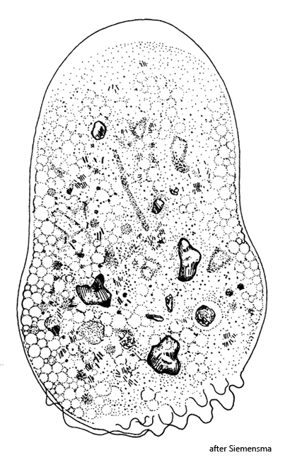
cytoplasma vacuolated, filled with detritus and mineral grains
up to several hundred nuclei
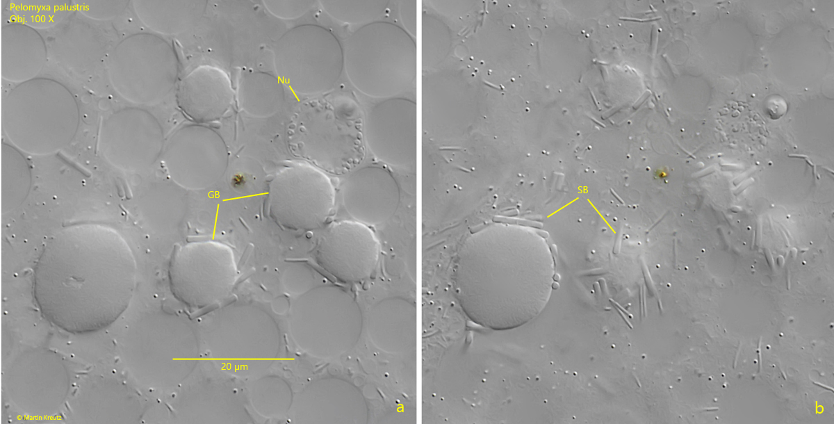
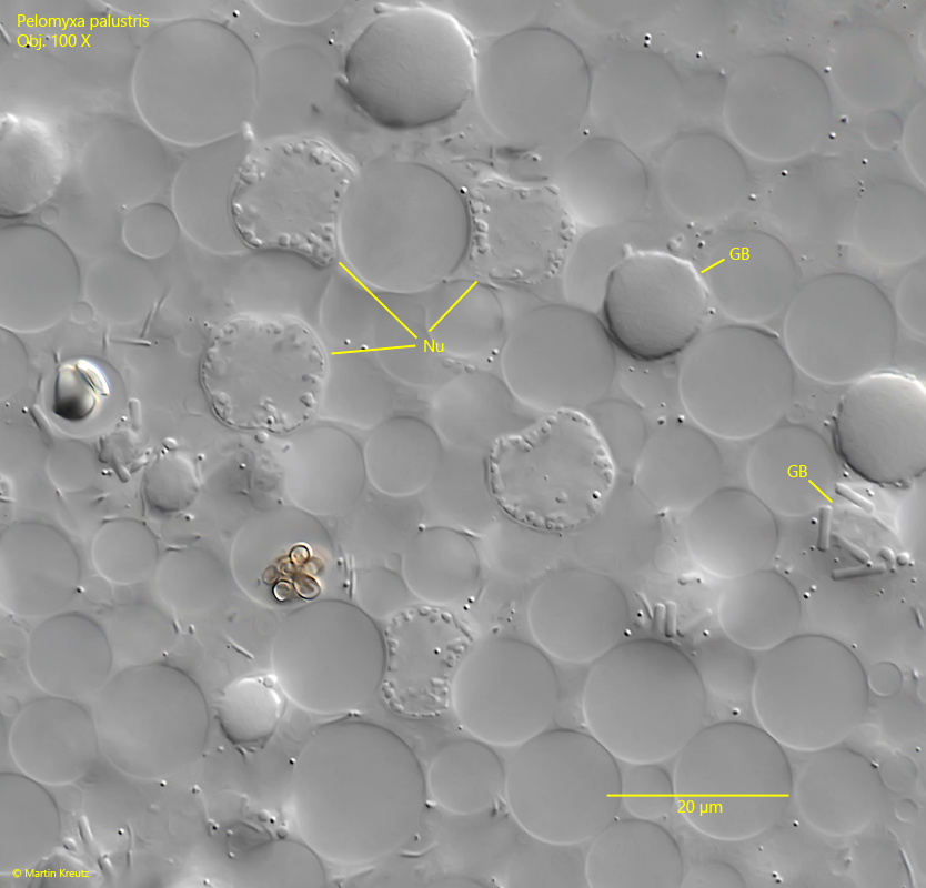
large, refractive glycogen bodies
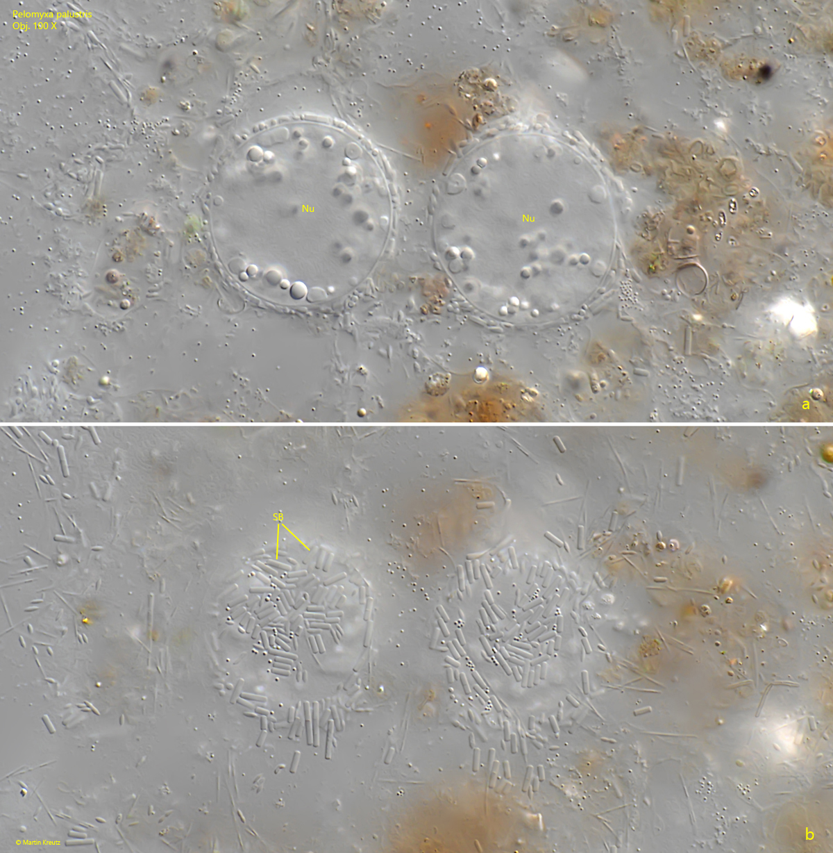
glycogend bodies covered with symbiotic bacteria
older nuclei covered by bacteria, young nuclei naked
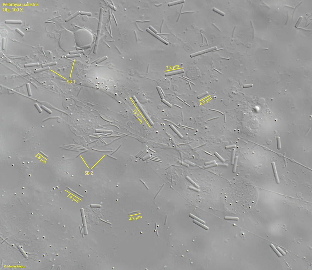
at least 2 species of symbiotic bacteria scattered in cytoplasm
temporarely flagella can be formed
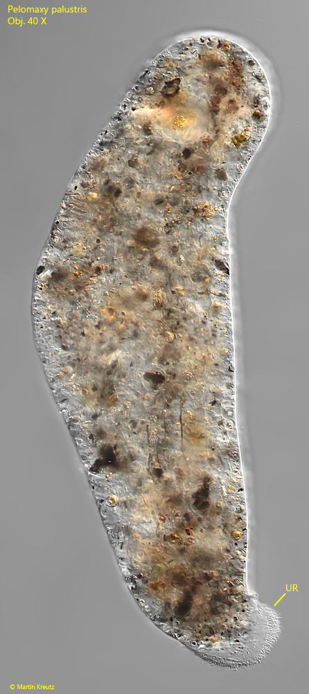
filose uroid is formed in monopodial movement
Pelomyxa palustris
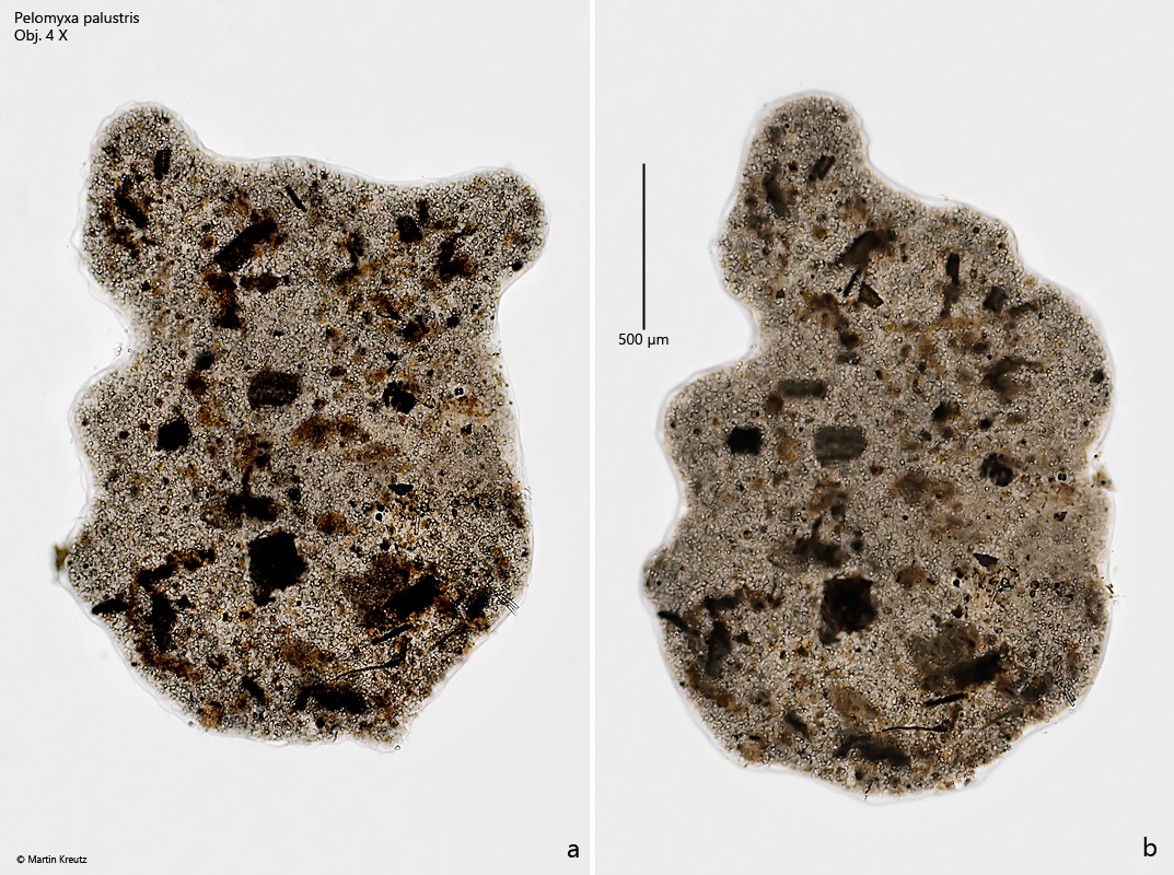
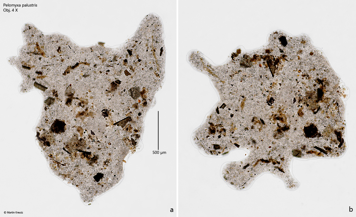
Pelomyxa palustris is one of the largest amoebas and is usually visible to the naked eye as gray-white or brownish-white bodies. I often find Pelomyxa palustris in locations with a thicker layer of mud, because Pelomyxa palustris is an anaerobic amoeba and lives only in the deeper layers.
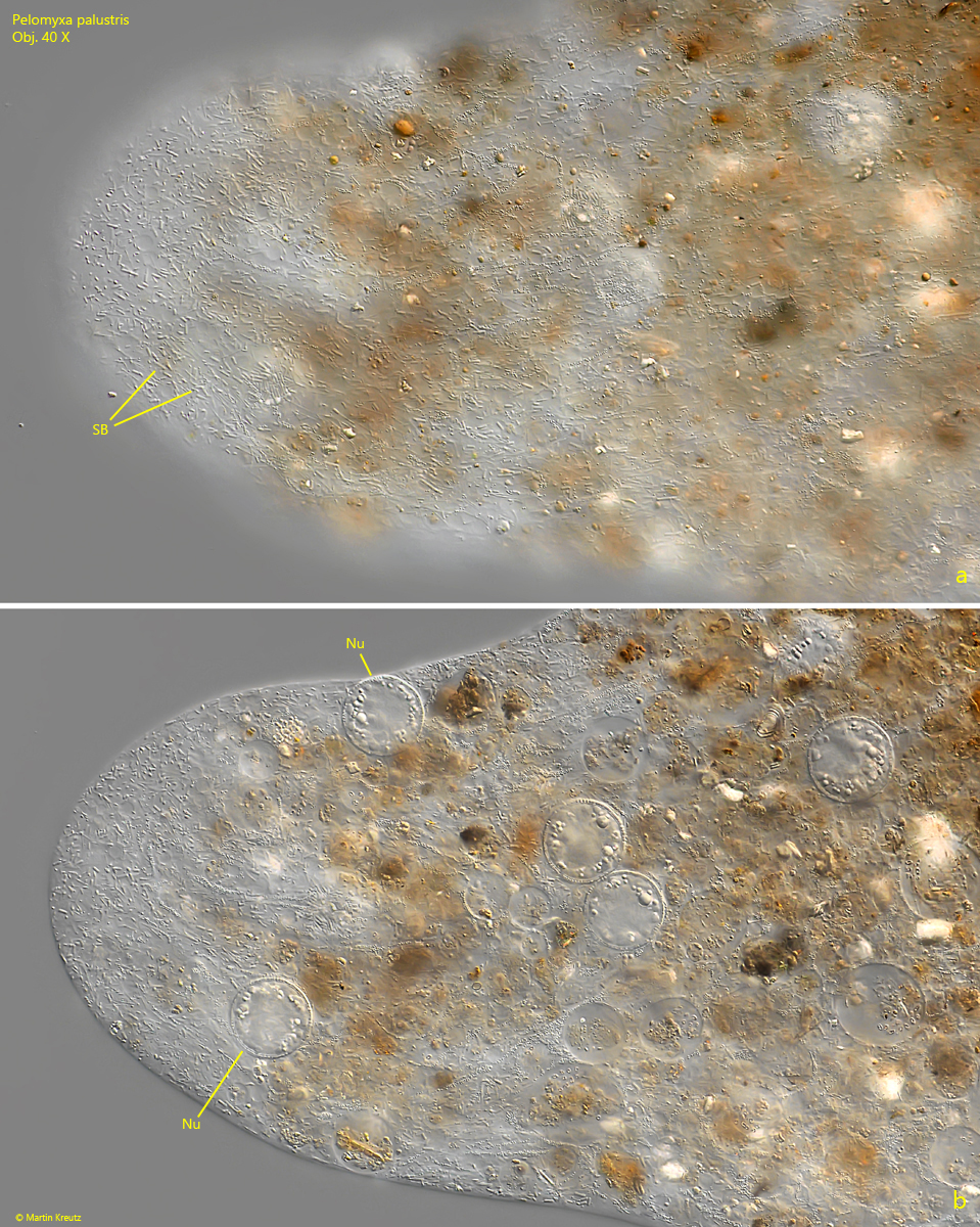
Under microscopic observation, the entire body is heavily filled with ingested detritus, mineral grains, algae, and fungal spores. As a result, Pelomyxa palustris is usually dark and translucent (s. figs. 1 a-b and 2 a-b). However, in squashed specimens, it can be seen that the cytoplasm is complexly structured. It is highly vacuolated and filled with numerous symbiotic bacteria (s. figs. 3 a and 8). In addition to the many nuclei scattered in the cytoplasm, numerous glycogen bodies can also be seen. The glycogen bodies are homogeneously structured and always covered with symbiotic bacteria (s. fig. 5 a-b). The young nuclei, on the other hand, are not always covered by bacteria (s. fig. 6 a-b), while older, larger nuclei are covered with the larger symbiotic bacteria of type 1 (s. fig. 7 a-b). The nuclei in Pelomyxa palustris have their own population dynamics. They can divide but also fuse into larger nuclei.
Pelomyxa palustris possesses neither mitochondria, Golgi apparatus, nor contractile vacuoles. The functions of these organelles are at least partially taken over by the symbiotic bacteria. The population dynamics of these bacteria within the cells and also after cell division are very complex and their details are not yet fully understood. However, it is certain that these are methanogenic bacteria, which enable the amoeba to live under anaerobic conditions.
Pelomyxa palustris can form short flagella, which can be distributed over the body; they are only short and obviously without function. Movement is not possible with these flagella, as they are immobile. The presence of these flagella is interpreted as a relic because Pelomyxa palustris evolved from flagellates. The flagella are formed only temporarily. I have not been able to observe them myself so far.
Fig. 1 a-b:Pelomyxa palustris. L = 2150 µm. A dark colored specimen filled up with ingested detritus particles. Obj. 4 X.
Fig. 2 a-b:Pelomyxa palustris. L = 2570 µm. A second, brighter specimen. Obj. 4 X.
Fig. 3 a-b:Pelomyxa palustris. Two focal planes of a pseudopodia. In the cytoplasm large amounts of symbiotic bacteria (SB) are visible und some large nuclei (Nu). Obj. 40 X.
Fig. 4:Pelomyxa palustris. L = 470 µm. A specimen in the monopodial form with a filose uroid (UR). Obj. 40 X.
Fig. 5 a-b:Pelomyxa palustris. Some of the glycogen bodies (GB) scattered in the cytoplasm. The diameter of the glycogen bodies is 5–15 µm und the content is homogeneous. All glycogen bodies are covered with symbiotic bacteria (SB). Nu = nucleus. Obj. 100 X.
Fig. 6:Pelomyxa palustris. Some of the nuclei (Nu) scattered in the cytoplasm. In this specimen, the nuclei are not covered with symbiotic bacteria. GB = glycogen bodies. Obj. 100 X.
Fig. 7 a-b:Pelomyxa palustris. Two large nuclei (Nu) with a diameter of 30 µm each. Both nuclei are covered with symbiotic bacteria (SB). Obj. 100 X.
Fig. 8:Pelomyxa palustris. In the cytoplasm at least 2 types of symbiotic bacteria are scattered. The type 1 bacteria (SB 1) are thick rods with a length of 4–15 µm, while the type 2 bacteria (SB 2) are thin rods with a length of 3-10 µm. Obj. 100 X.