cell in lorica with transverse ridges, narrow aperture
length (of cell) about 100 µm
length of lorica about 150 µm
oral aperture apical, surrounded by rows of cilia
somatic cilia in transverse rows
pellicle with rectangular pattern
cytoplasm pink due to ingested purple bacteria
contractile vacuole near mid-body
macronucleus globular with adjacent micronucleus
several caudal cilia
Vasicola ciliata
I find Vasicola ciliata very frequently in my sampling sites. The species is anaerobic and lives in the deeper mud layers. The populations vary greatly, as they also correlate with the presence of purple bacteria, on which Vasicola ciliata mainly feeds. As a result, the specimens are always strongly pink-colored and opaque.
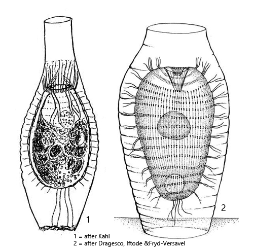
Vasicola ciliata builds its own lorica in which the ciliate lives and draws in food. The lorica is a swollen tube with a narrowed, apical opening. The middle part of the lorica has transverse ridges, which appear as a wave pattern in optical section. The lorica is usually attached to detritus flakes. It is thin-walled, transparent, and colorless. The ciliate inside the lorica usually takes on a broadly ellipsoid shape, and the somatic cilia grip the transverse ridges, providing it with support (s. figs. 2 and 3).
In the samples, one often also finds freely swimming specimens because Vasicola ciliata quickly leaves the lorica when disturbed. This is especially the case after placing the coverslip. In the freely swimming specimens, the caudal cilia can be seen quite well (s. fig. 7 a), which are difficult to recognize inside the lorica.
The pellicle of Vasicola ciliata shows a typical and very regular, rectangular pattern (s. figs. 7 b, 8 and 9). It connects the rows of somatic cilia, which are distributed in straight transverse rows over the body (s. fig. 10 a). In the front part of the body, an alveolar system of canals can also be seen under the pellicle (s. fig. 10 b). However, I could not recognize these canals in all specimens.
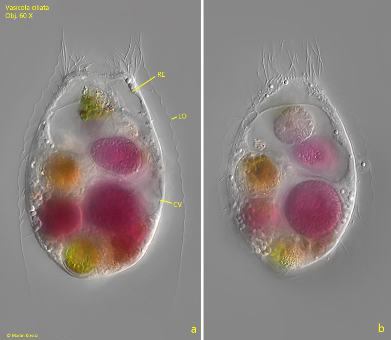
Fig. 1 a-b:Vasicola ciliata. L = 95 µm (of cell). Two focal planes of a specimen in the lorica (LO). In the apical fourth the cavity below the oral aperture is visible, called receptaculum (RE). In the cytoplasm pink and greenish food vacuoles are visible from ingested purple bacteria and algae. CV = contractile vacuole. Ob. 60 X.
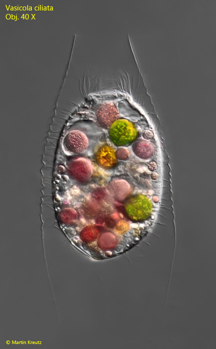
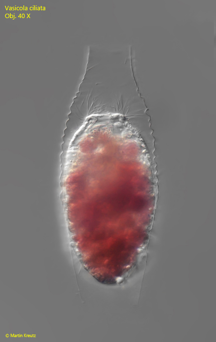
Fig. 2:Vasicola ciliata. L = 92 µm (of cell). A specimen found in November 1998 in the Simmelried. Ob. 40 X.
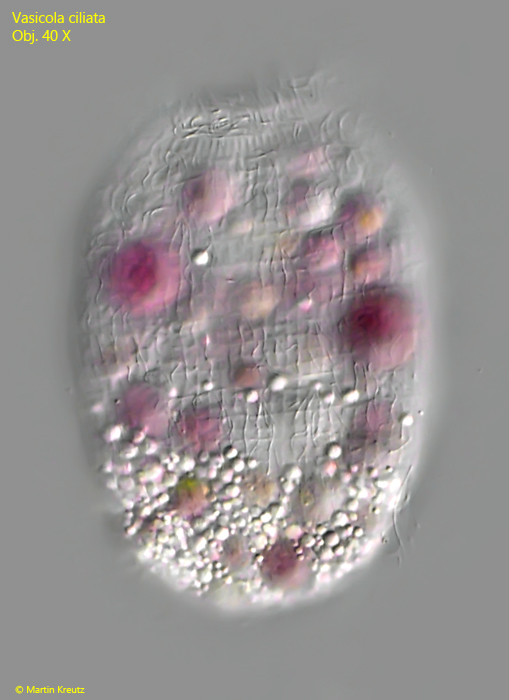
Fig. 3:Vasicola ciliata. L = 108 µm (of cell). A specimen found in June 2022 in the Simmelried. Ob. 40 X.
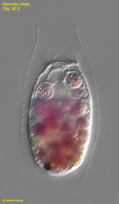
Fig. 4:Vasicola ciliata. L = 124 µm (of cell). A well-nourished specimen found in January 2014 in the Ulmisried. Ob. 40 X.
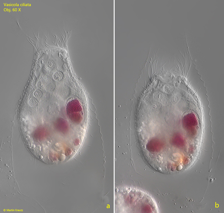
Fig. 5 a-b:Vasicola ciliata. L = 81 µm (of cell). A specimen found in January 2013 in the Simmelried. Ob. 60 X.
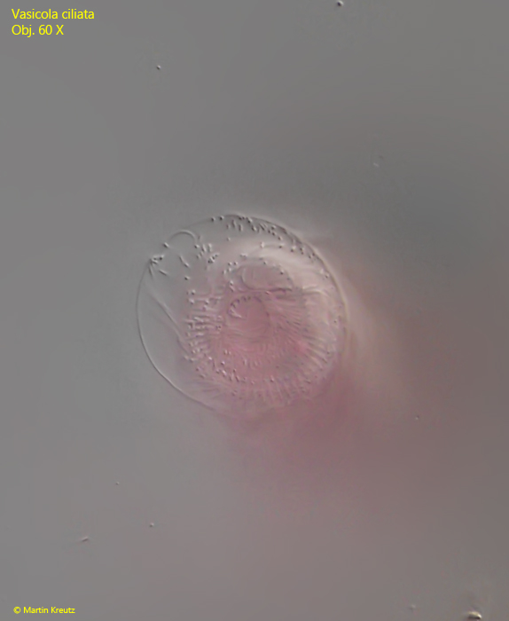
Fig. 6:Vasicola ciliata. Apical view on the oral aperture of a specimen in the lorica. Ob. 60 X.
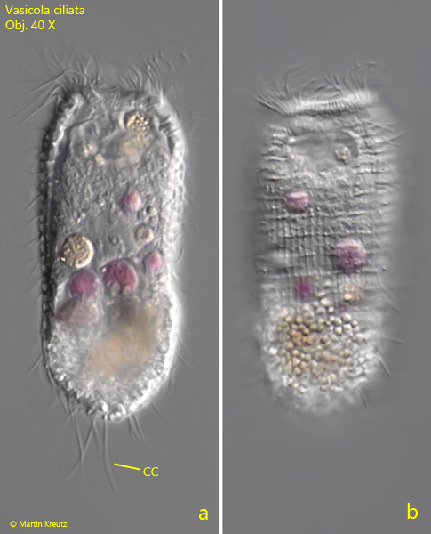
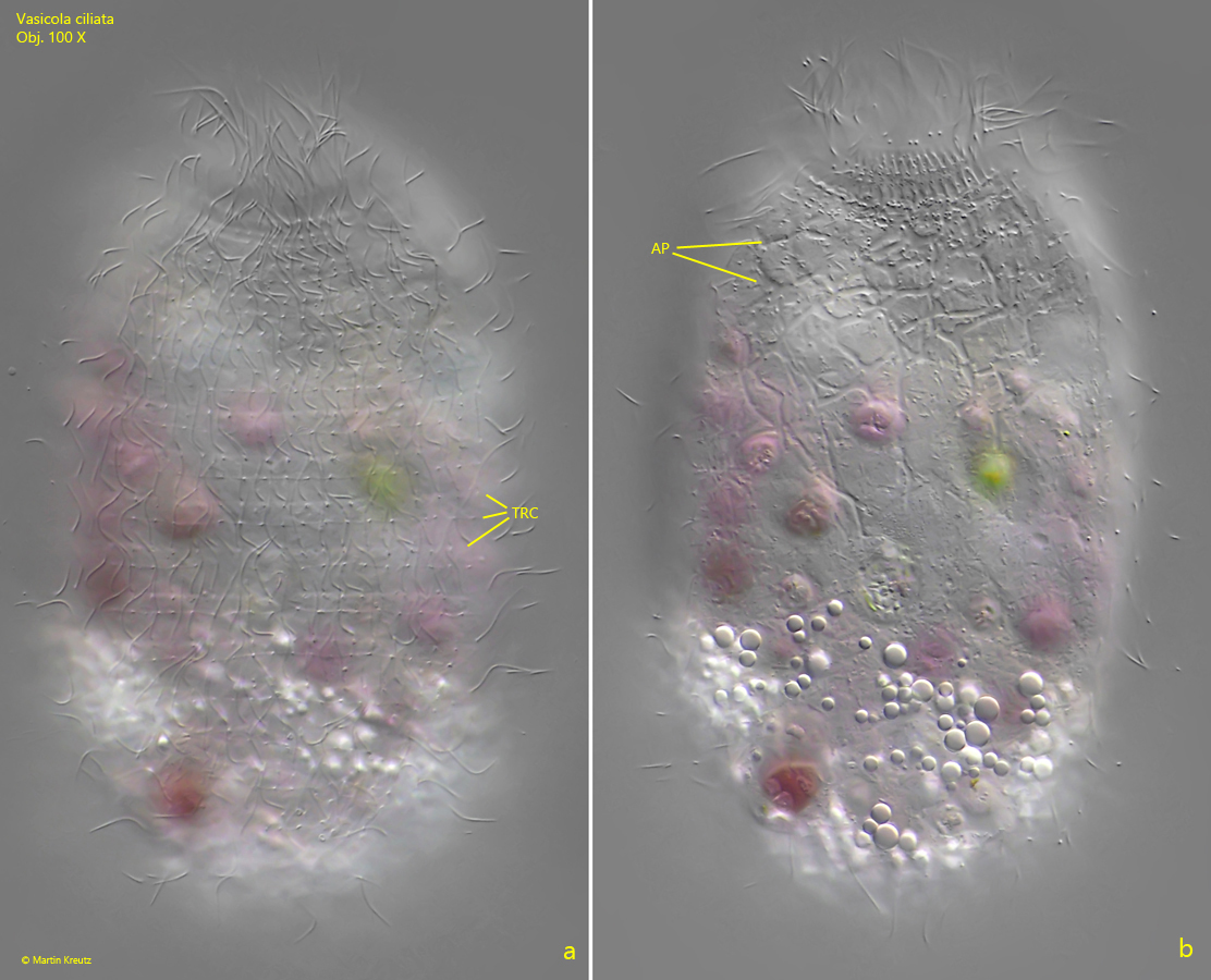
Fig. 7 a-b:Vasicola ciliata. L = 90 µm. two focal planes of a freely swimming specimen found in November 1997 in the Simmelried. Note the caudal cilia (CC) and the reticulate pattern of the pellicle (b). Ob. 40 X.
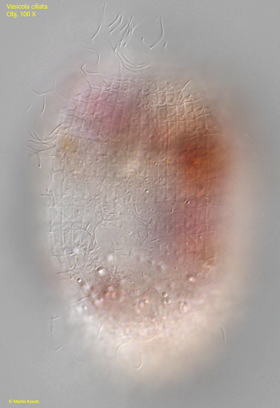
Fig. 8:Vasicola ciliata. The reticulate pattern of the pellicle in a slightly squashed specimen. Ob. 40 X.
Fig. 9:Vasicola ciliata. The reticulate pattern of the pellicle in a squashed specimen. Ob. 100 X.
Fig. 10 a-b:Vasicola ciliata. Focal plane on the transverse rows of somatic cilia (TRC) and on the canal-shaped alveolar pattern (AP) in the anterior third of the body. Ob. 100 X.
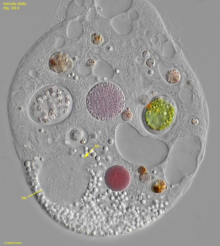
Fig. 11:Vasicola ciliata. The macronucleus (Ma) and micronucleus (Mi) in a strongly squashed specimen. Ob. 100 X.