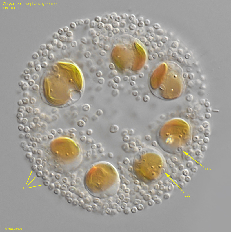
numerous excretion bodies scattered in gelatinous sheath
cells globular, young cells amoeboid
length 8.5–10 µm, width 6.5–8 µm (of cells)
cells extending fine filopodia from colony
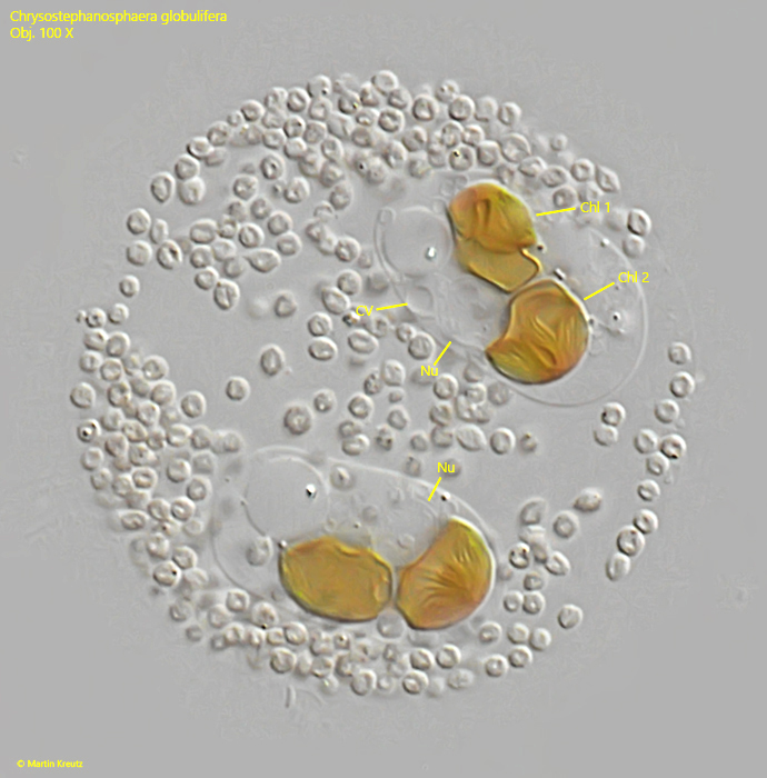
two chloroplasts per cell
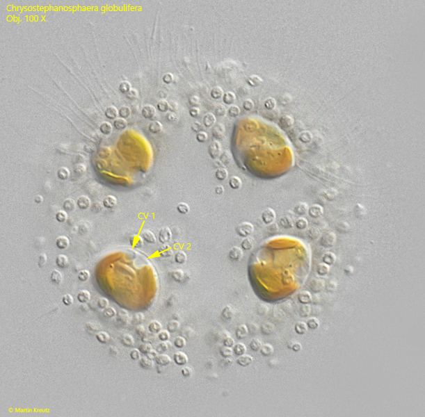
two contractile vacuoles posterior
spherical nucleus between chloroplasts
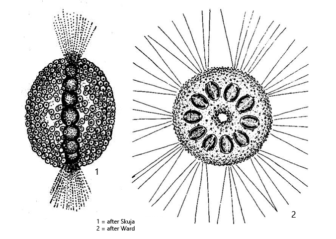
Chrysostephanosphaera globulifera
I have found Chrysostephanosphaera globulifera in large quantities in samples from the Schwemm Moor in Austria. In my local area, I find Chrysostephanosphaera globulifera exclusively in the Simmelried and there only very rarely.
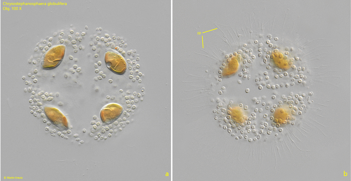
The disc-shaped colonies of Chrysostephanosphaera globulifera are very characteristic due to the circular arrangement of the cells and the many spherical excretion bodies with which the gelatinous envelope of the colonies is interspersed. In addition, fine filopodia of the cells can often be observed, which extend radially from the colonies (s. fig. 2 a-b). These are very fine and can only be seen at high magnification. According to my observations, these filopodia are particularly noticeable in young colonies, in which the cells are still amoeboid or elliptically shaped. The older cells round up and only rarely show these filopodia.
There were different theories from earlier authors regarding the nature and origin of the excretion bodies. They were considered symbiotic bacteria by Geitler (1948), Starmach (1985), and John et al. (2002). Stein (1858) and Scherffel (1911) regarded them as excretion bodies. Laber (2019) was able to show through autofluorescence that they cannot be cyanobacteria either. Instead, he demonstrated that they are endogenously produced excretion bodies, which are produced and secreted by the cells themselves. (s. link “Mikro-Forum” below). I can confirm Laber’s results, as the excretion bodies not yet secreted could be clearly seen in separate vacuoles within the cells (s. fig. 6).
The cells of Chrysostephanosphaera globulifera have two chloroplasts and two contractile vacuoles. The two chloroplasts are only visible in heavily squashed cells (s. fig. 4). They surround the nucleus. The two contractile vacuoles are rarely seen at the same time (s. fig. 5). According to my observations, they can be located in the inward-facing part of the cell, but also on the outward-facing side. This may depend on the age of the cells.
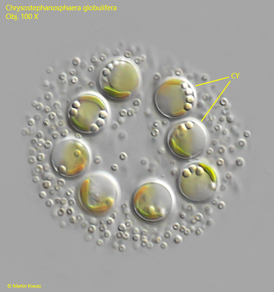
Reproduction occurs through flagellated swarmers. These transform into amoeboid stages, which begin to build a new colony (s. fig. 8). In older colonies, I was able to observe spherical cysts with a distinct cell wall on one occasion, which may serve as resting stages for overwintering (s. fig. 9).
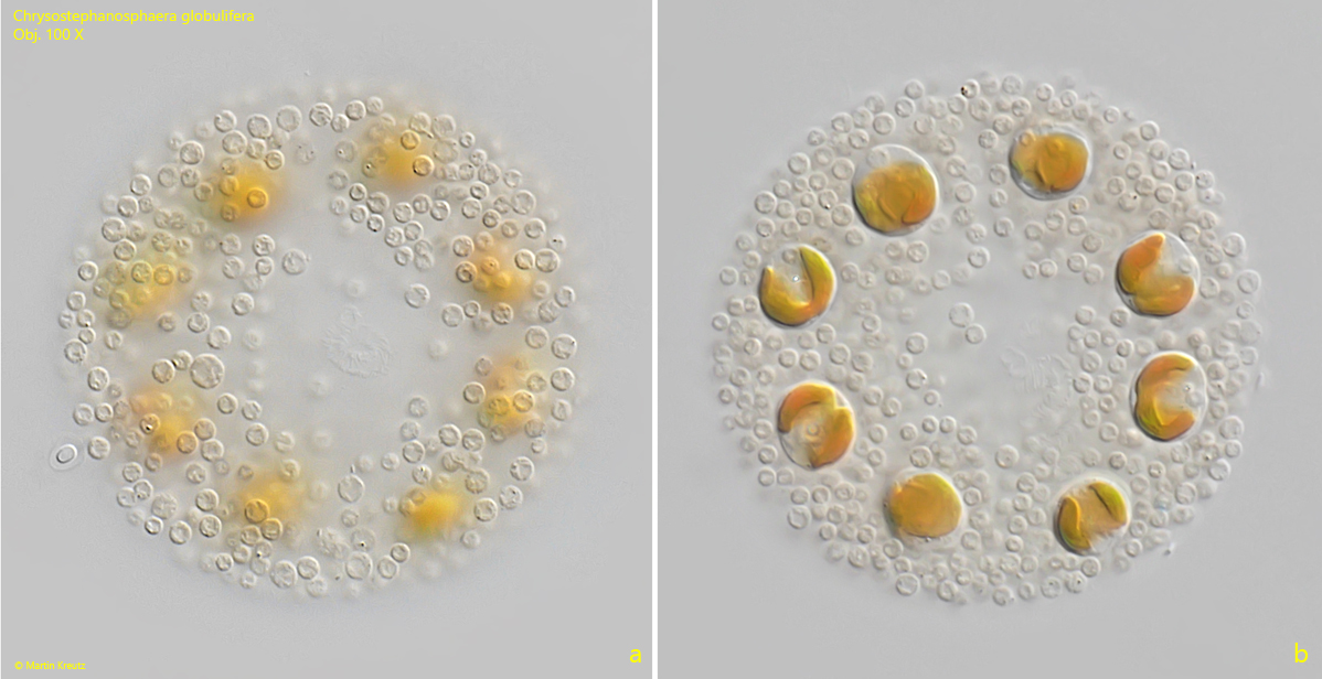
Fig. 1 a-b:Chrysostephanosphaera globulifera. D = 70 µm (of colony). Two focal planes of a slightly squashed colony of eight cells. The almost spherical cells have a diameter of 10–11 µm. Obj. 100 X.
Fig. 2 a-b:Chrysostephanosphaera globulifera. D = 63 µm (of colony). Two focal planes of a young colony of four, ellipsoid cells. Note the delicate filopodia (FP) extendings radially from the colony. Obj. 100 X.
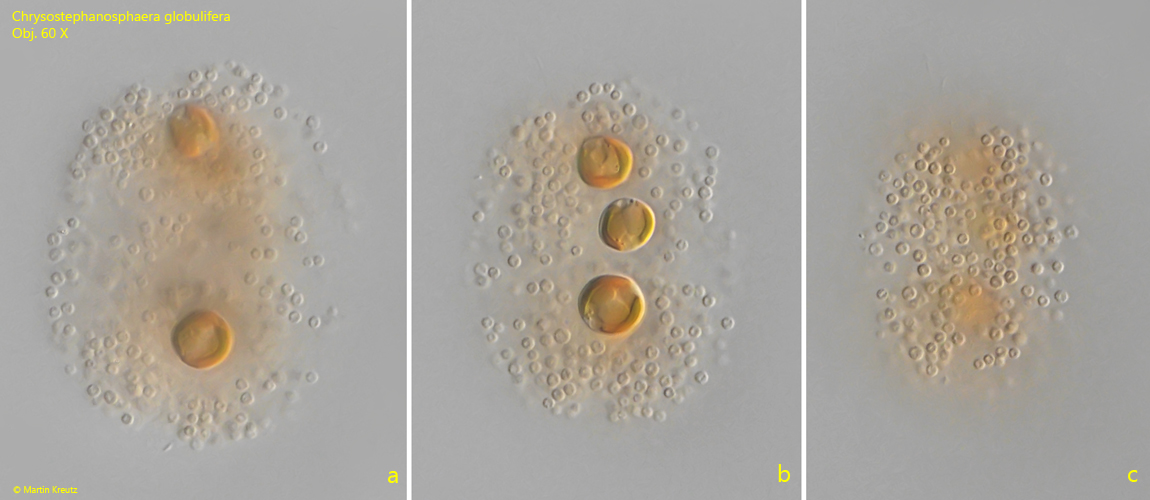
Fig. 3 a-c:Chrysostephanosphaera globulifera. D = 65 µm (of colony). Three focal planes of a colony in lateral view. Obj. 60 X.
Fig. 4:Chrysostephanosphaera globulifera. A strongly squashed colony of two cells for visualization of the two chloroplasts per cell (Chl 1, Chl 2) and the nucleus (Nu) located between the chloroplasts. CV = contractile vacuole. Obj. 100 X.
Fig. 5:Chrysostephanosphaera globulifera. A slightly squashed colony. In one cell the two contractile vacuoles (CV) are visible. Obj. 100 X.
Fig. 6:Chrysostephanosphaera globulifera. The spherical excretion bodies (EB) are produced and excreted by the cells. In two of the cells the endogenously produced excretion bodies (EEB) can be seen enclosed in separate vacuoles. The excretion bodies have a diameter of 1.5–2.0 µm. Obj. 100 X.
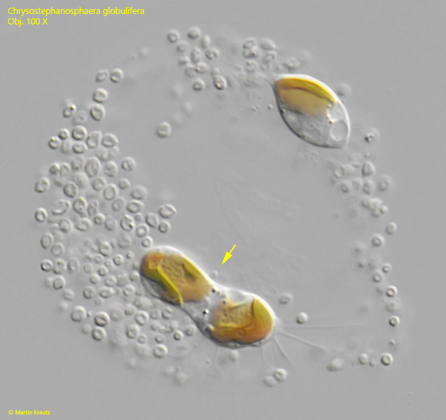
Fig. 7:Chrysostephanosphaera globulifera. A cell division (arrow) in a young colony of only two cells. Obj. 100 X.
Fig. 8:Chrysostephanosphaera globulifera. An amoeboid cell with numerous filopodia starts to create a new colony. Nu = nucleus, CV = contractile vacuole. Obj. 100 X.
Fig. 9: Chrysostephanosphaera globulifera. D = 38 µm (of colony). A colony with eight spherical cysts (CY) with a distinct cell wall. Obj. 100 X.