Nassula ornata Ehrenberg, 1833
Most likely ID: n.a.
Â
Synonym: n.a.
Â
Sampling location: Main bike path pond
Â
Phylogenetic tree: Nassula ornataÂ
Â
Diagnosis:Â
- body broad ellipsoidal, anterior end somewhat broader
- length 155–320 µm, width 100–200 µm
- oral basket in anterior third, 20–32 nematodesmal rods
- basket with inconspicuous ring structure
- cells colorful due to colored food vacuoles
- synhymenium starts below basket, run to dorsal side
- spot of orange-yellow granules dorsally
- contractile vacuole central on ventrsal side, one excretion pore
- macronucleus spherical with 2–14 adjacent micronuclei
- extrusomes spindle-shaped, 8–10 µm long
- 140–190 longitudinal rows of cilia
In my immediate surroundings, I have never been able to detect Nassula ornata. It was only in March 2026 that I was able to observe a mass development of Nassula ornata in a shallow pond next to the Main bike path near Würzburg. The pond appears to be heavily eutrophic with an anaerobic, black sludge layer at a depth of 10–15 cm. This sludge layer is covered by extensive mats of cyanobacteria, mainly of the genus Oscillatoria.
Â
Nassula ornata is one of the most colorful ciliates overall. The cytoplasm is densely filled with many food vacuoles of different colors. Violet and orange tones dominate, but blue and green food vacuoles can also be observed. The colors can be very intense (s. figs. 4 a-b, 5 a-b and 6 a-b). The colors result from the phagocytized cyanobacteria, on which Nassula ornata feeds exclusively. During the digestive process, the color of the food vacuoles changes. Additionally, Nassula ornata has a yellow-orange spot on the “neck,” which can vary in intensity (s. fig. 11). Because of this play of colors, Nassula ornata is a photogenic subject.
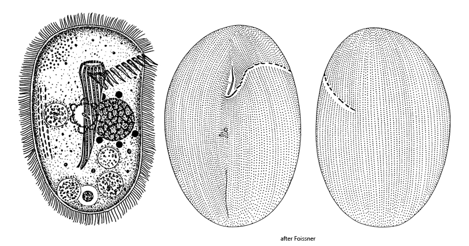
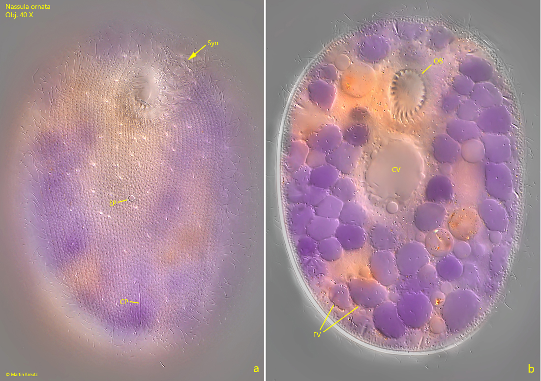
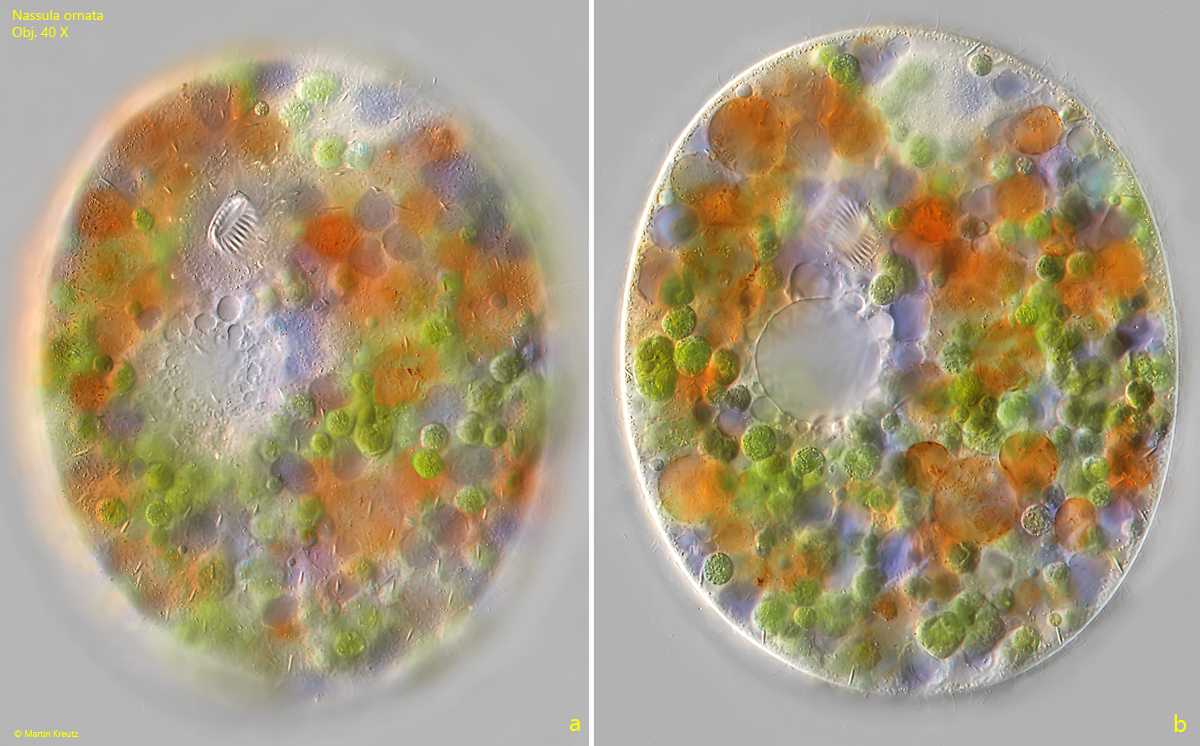
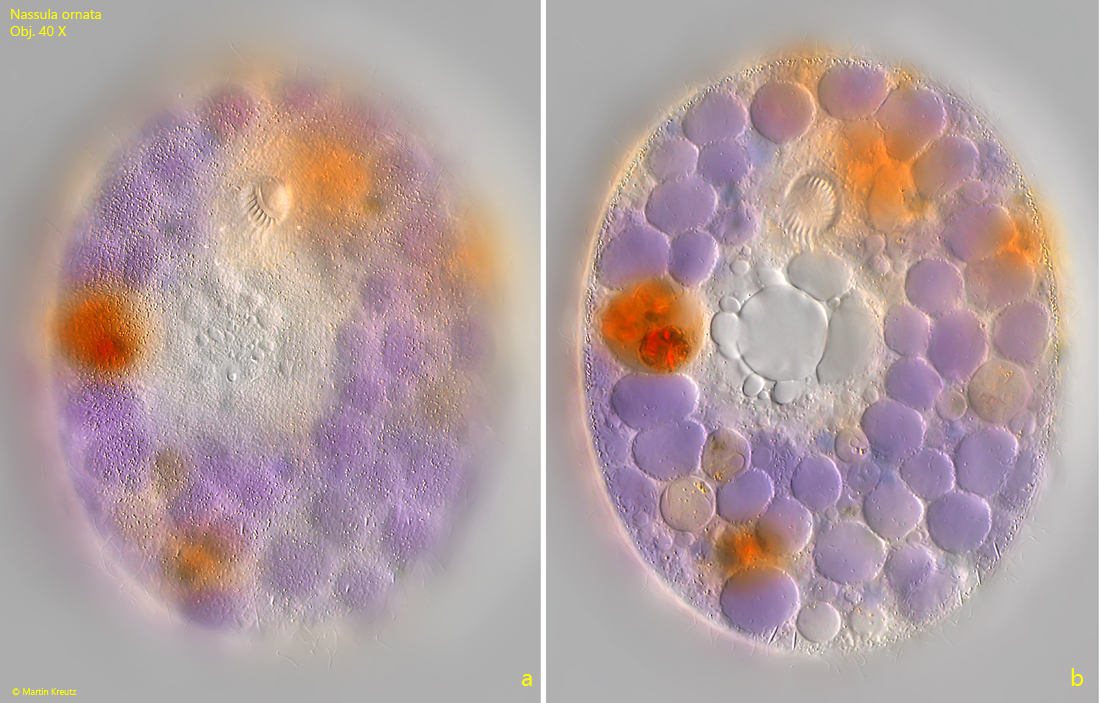
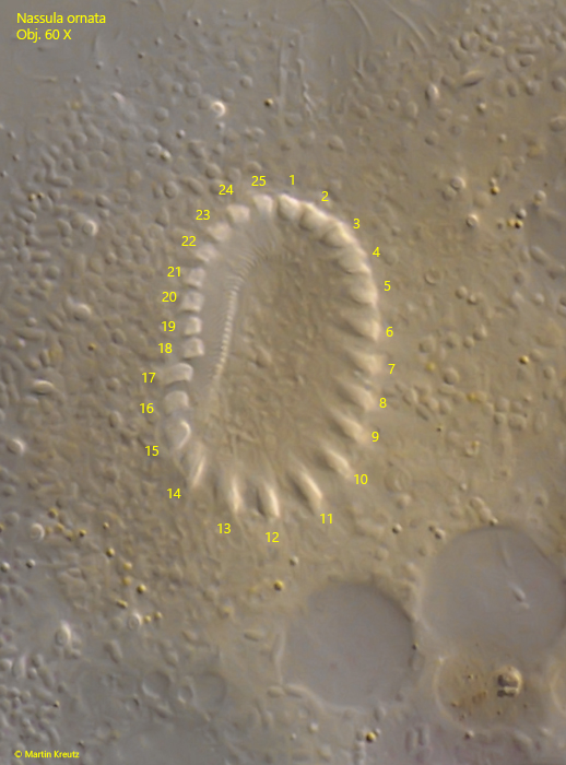
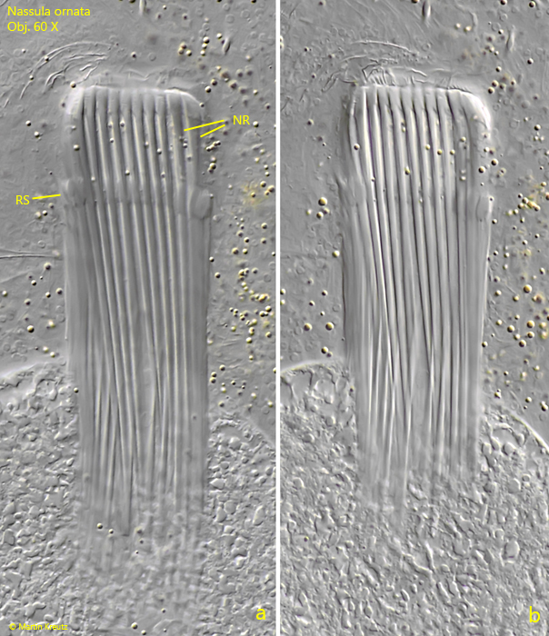
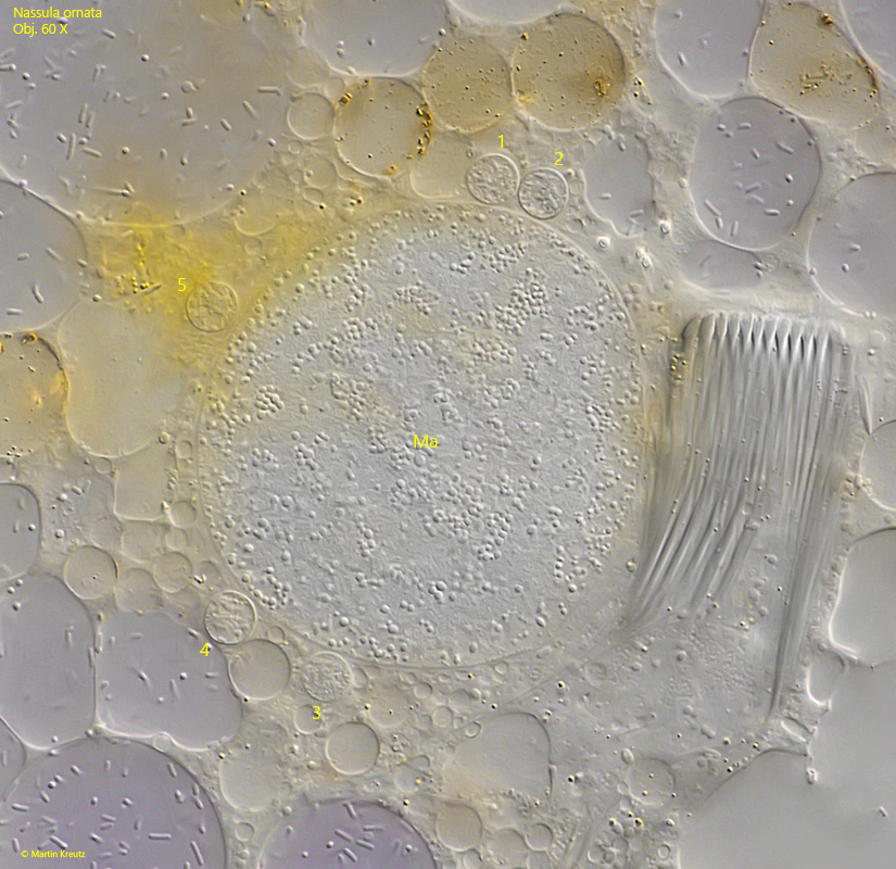
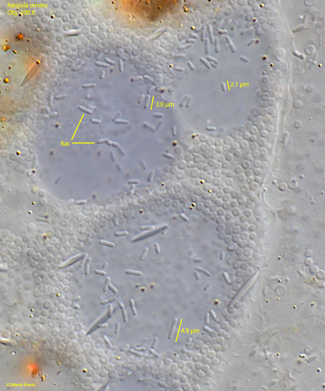
Nassula ornata primarily feeds on filamentous cyanobacteria such as Oscillatoria, Phormidium, or Anabaena. The filaments are phagocytosed with the help of the oral basket. This consists of 20–32 nematodesmal rods that can slide against each other. This allows the oral basket to adjust to the thickness of the ingested cyanobacteria. In strongly squashed specimens, a ring structure can be seen near the distal end of the oral basket (s. fig. 8), which, however, is not as pronounced as in the similar species Obertrumia aurea.
Â
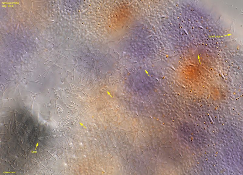
As a nassulid ciliate, Nassula ornata possesses a so-called synhymenium. This is a band of ciliary tufts that begins below the oral basket on the ventral side, runs over the left side, and finally ends on the dorsal side. The ciliary tufts are particularly difficult to recognize in compressed specimens because they are pressed against the body. Therefore, the basal bodies of the ciliary tufts are focused on in order to recognize the synhymenium (s. figs. 9 and 10). The shape and length of the synhymenium is an important distinguishing feature between the genera within the nassulid ciliates (s. also Obertrumia aurea). In Nassula ornata, the synhymenium has no interruptions and runs slightly S-shaped.
The specimens of my population were mostly between 220-300 µm long. The length can vary greatly depending on nutrition. The globular macronucleus is located centrally, behind the contractile vacuole, which is on the ventral side and has a single excretory pore (s. fig. 4 a-b). In my specimens, the macronucleus was usually accompanied by 5–6 spherical micronuclei. Scattered under the pellicle, numerous spindle-shaped extrusomes can be seen, which in my population were 10–11.5 µm long (s. fig. 13).
Â
In squashed specimens, I examined the colored food vacuoles more closely. I found that bacteria were always present especially in the violet-colored vacuoles with clear content, whereas no bacteria were found in the orange or yellow food vacuoles, which also contained numerous particles (s. fig. 14). One might assume that the bacteria are taken up together with the phagocytosed cyanobacteria. However, in that case, one would expect different species to be found in the violet food vacuoles. This is not the case, however, because the same type of bacteria is found in all vacuoles. They are 2–5 µm long rods with rounded ends (s. fig. 15). I could not find any explanation for these bacteria in the literature. Possibly, they are a type of symbiont that helps in the decomposition of food.
Â
Nassula ornata is phototactically negative (Raikov, 1962). I was able to observe this behavior very clearly in one of my sample jars. Several thousand specimens had gathered near the surface. When I illuminated them with a strong flashlight, it was possible to see with the naked eye how the specimens quickly moved to deeper layers and avoided the light. Which organelle is responsible for the photosensitivity remains unclear.
Â
Nassula ornata can easily be confused with the similar species Obertrumia aurea. This species can also form colored food vacuoles, which are usually yellow or orange. However, Obertrumia aurea does not have extrusomes, and the synhymenium on the dorsal side abruptly transitions from an S-shaped course to a straight line. Additionally, Obertrumia aurea has a pronounced ring structure on the oral basket, which is easy to recognize even in an uncompressed specimen.
Fig. 1: Nassula ornata. L = 180 µm. Several freely swimming specimens in brightfield illumination. Obj. 100 X.
Fig. 2 a-c: Nassula ornata. 235 µm. A freely swimming specimen from ventral (a, b) and from left (c). Obj. 40 X.
Fig. 3 a-c: Nassula ornata. 283 µm. A second freely swimming specimen from ventral (a, b) and from left (c). Obj. 40 X.
Fig. 4 a-b: Nassula ornata. 240 µm. Two focal planes of a slightly squashed specimen from ventral. CV = contractile vacuole, CP = slit-shaped cytoproct, EP = excretion pore of contractile vacule, FV = food vacuoles, OB = oral basket, Syn = synhymenium. Obj. 60 X.
Fig. 5 a-b: Nassula ornata. 258 µm. Two focal planes of a second, slightly squashed specimen from ventral. Due to freshly ingested cyanobacteria (Oscillatoria spec.) many food vacuoles are colored green. Obj. 40 X.
Fig. 6 a-b: Nassula ornata. 234 µm. Two focal planes of a third, slightly squashed specimen from ventral. Obj. 40 X.
Fig. 7: Nassula ornata. The oral basket in ventral view. In this specimen it constists of 25 nematodesmal rods (1–25). Obj. 60 X.
Fig. 8: Nassula ornata. The oral basket in a strongly squashed specimen. Near the anterior end of the nematodesmal rods (NR) a characteristic ring structure (RS) is visible. Obj. 60 X.
Fig. 9: Nassula ornata. The synhymenium (arrows) starts below the oral basket on the ventral side and run to the left side. It ends on the dorsal side. Obj. 100 X.
Fig. 10: Nassula ornata. In a strongly squashed specimen the course of the synhymenium (arrows) over the left side to the end on the dorsal side is visible. Obj. 100 X.
Fig. 11: Nassula ornata. At the height of the oral basket, there is a yellow-orange spot on the dorsal side. It consists of a dense cluster of strongly colored vesicles. Obj. 60 X.
Fig. 12: Nassula ornata. The globular macronucleus (Ma) with 5 adjacent, spherical micronulei (1–5). Obj. 100 X.
Fig. 13: Nassula ornata. The extrusomes (EX) are spindle-shaped and have a length of 10.5–11.5 µm. Obj. 100 X.
Fig. 14: Nassula ornata. Some of the different colored food vacuoles (FV) in detail. Note that exclusively in the violet colored food vacuoles bacteria (Bac) are present. Obj. 100 X.
Fig. 15: Nassula ornata. The bacteria (Bac) in the violet food vacuoles are from the same type with a length of 2–5 µm. Obj. 100 X.
Fig. 16: Nassula ornata. 260 + 280 µm. Two specimens in the process of conjugation. Obj. 40 X.