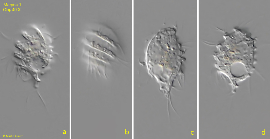
In May 2021, I found a small ciliate with a length of 25–30 µm in a moss sample from a tree. The shape of the ciliate resembled a pine cone. The anterior two-thirds of the body was approximately helmet-shaped. The posterior third tapered in a cone shape. A spiral ridge ran over the entire body in 7–8 turns counterclockwise to the posterior end (s. figs. 1 a-d and fig. 3 a-h). This ridge showed knob-like protuberances (s. fig. 3 a). At the posterior end, I could identify at least two caudal cilia (s. fig. 1 c). The somatic cilia always arose in pairs. The mouth opening was located at the posterior end, where the conical tapering begins. At the same level was also the contractile vacuole located. The round macronucleus was always located in the anterior half of the body, together with a small, attached micronucleus. I always found the specimens of this ciliate freely swimming, where they rotated around their longitudinal axis.
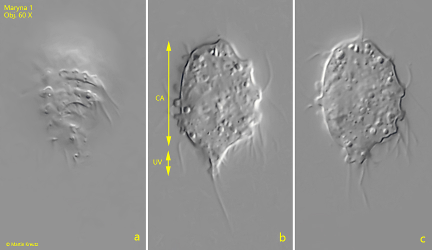
The sum of these characteristics pointed to the genus Maryna, a genus within the Colpodida. The genus Maryna is characterized by a helmet-shaped front part of the body, which is called the calix (s. fig. 2 b). The postoral part of the body is greatly reduced and often dome-shaped, cone-shaped, or cylindrical. This part of the body is called the uvula (s. fig. 2 b).
All species within the genus Maryna build gelatinous loricae, which are mostly tubular. The ciliates then sit at the end of these tubes, with the uvula extended out of the tube, while the calix remains inside the tube. Although I found all my specimens to be free-swimming, it is not excluded that this species also builds loricae, as these are abandoned at the slightest disturbance. Since I sucked my specimens out of the moistened moss with a pipette, it was unlikely to find specimens inside a lorica.
Within the genus Maryna, I have not been able to find any species with the characteristics described above. It is therefore not excluded that this is a previously undescribed species Maryna nov. spec., which I provisionally designate as Maryna 1.
Fig. 1 a-d:Maryna 1. L = 27 µm. Different focal planes of a freely swimming specimen. Note the distinct spiral ridge with several counterclockwise turns. Obj. 40 X.
Fig. 2 a-c:Maryna 1. L = 25 µm. A second freely swimming specimen. The helmet-shaped anterior part is called the calix (CA) while the tapered posterior end is called uvula (UV). Obj. 60 X.
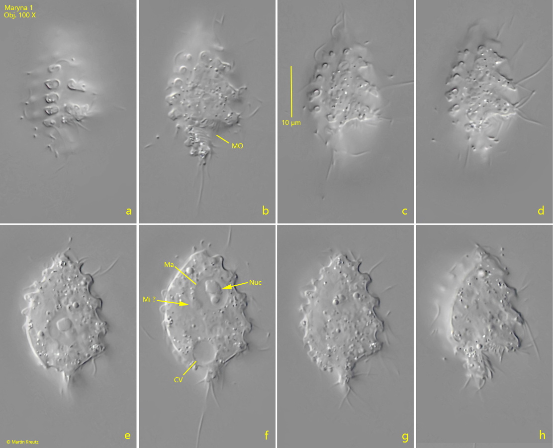
Fig. 3 a-h:Maryna 1. L = 28 µm. Different focal planes of a third specimen. The spiral ridge of this specimen has knob-shaped protuberances. The mouth opening (MO) is located posterior as well as the contractile vacuole (CV). The spherical macronucleus (Ma) has a central nucleolus (Nuc). Mi ? = probably the micronucleus. Obj. 100 X.
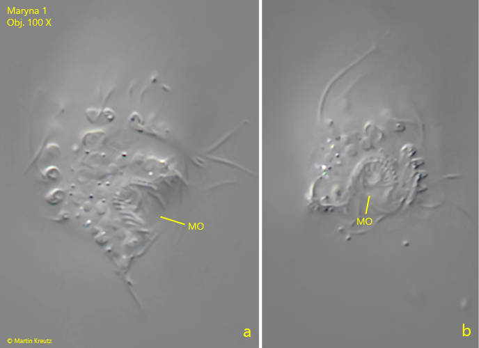
Fig. 4 a-b:Maryna 1. The mouth opening (MO) in lateral (a) and in posterior view (b). Obj. 100 X.