Uronema nigricans

(Müller, 1786) Florentin, 1901

Most likely ID: n.a.


Synonyms: Cyclidium nigricans, Uronema marinum, Uronema parduczi


Sampling location: Simmelried


Phylogenetic tree: Uronema nigricans


Diagnosis: 

  • body oval or barrel-shaped
  • length 25–50 µm, width 15–25 µm
  • dorsal side convex, oral apparatus slightly indented
  • obliquely truncated frontal plate
  • oral apparatus with 3 membranelles in anterior third
  • undulating membrane on right side
  • macronucleus spherical, one spherical micronucleus
  • contractile vacuole terminal
  • extrusomes rod-shaped, inconspicuous
  • cytoplasm often darkly spotted due to crystalline inclusions
  • 10–14 longitudinal rows of cilia
  • paired cilia in anterior third
  • one caudal cilium, half body length
Uronema-nigricans
Uronema nigricans

So far, I have found Uronema nigricans only a few times. Since the ciliate is quite small, I may have overlooked it more often, as this species is described as very common, especially in eutrophic locations.

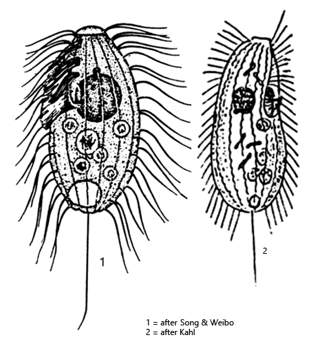
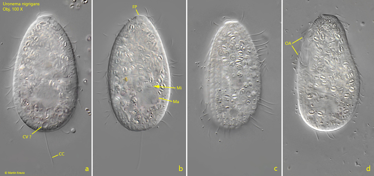
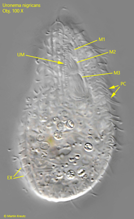
At high magnification, Uronema nigricans stands out due to its distinct frontal plate and the slightly ribbed pellicle (s. fig. 1 c). A reliable identification feature is the structure of the oral apparatus, which is, however, difficult to observe because Uronema nigricans is motile and hard to position ventrally. Then, one can recognize three adoral membranelles arranged one below the other and a right-sided undulating membrane (s. fig. 2). Another important feature is the paired cilia in the anterior part of the body. The posterior cilia stand individually. Since all my specimens were unfortunately filled with food, I could hardly discern the internal structure. A lens-shaped micronucleus is attached to the round macronucleus (s. fig. 1 b). I could not clearly identify the contractile vacuole. The caudal cilium (s. fig. 1 a) is only half the body length, as described by Foissner, Berger & Kohmann (1994).
Uronema-nigricans

Fig. 1 a-d: Uronema nigricans. L = 42 µm. A freely swimming, well-nourished specimen. CC = caudal cilium, CV = probably the contractile vacuole,  FP = frontal plate, Ma = macronucleus, Mi = micronucleus, OA = oral apparatus. Obj. 100 X.

Uronema-nigricans

Fig. 2: Uronema nigricans. L = 42 µm. The same specimen as shown in fig. 1 a-d in ventral view. Note the three adoral membranelles (M1–M3) of the oral apparatus with the undulating membrane (UM) on the right side. A part of the the somatic cilia are paired (PC). EX = extrusomes. Obj. 100 X.