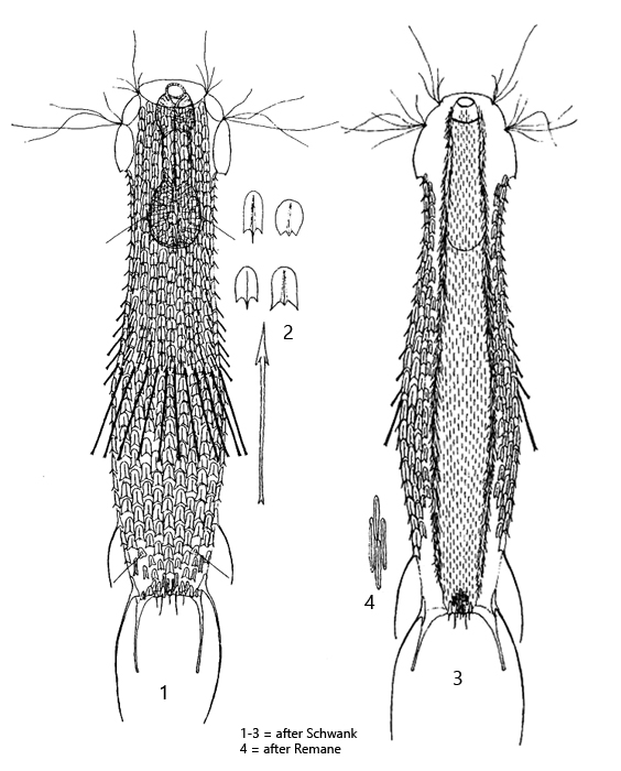
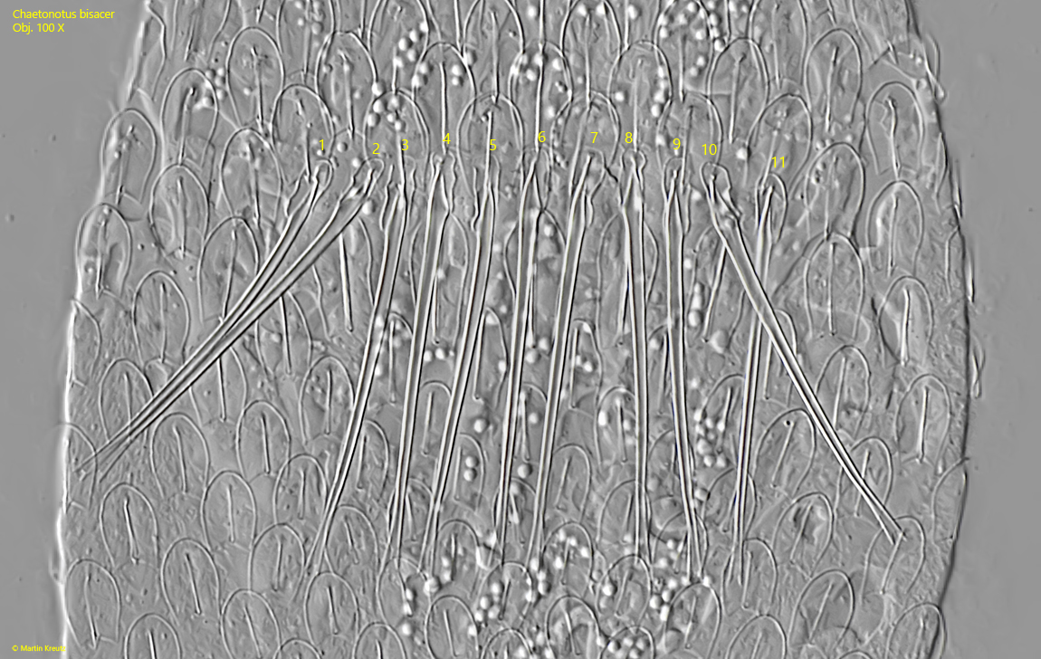
dorsal girdle of 7–11 (20) rod-shaped spines, double tip distally
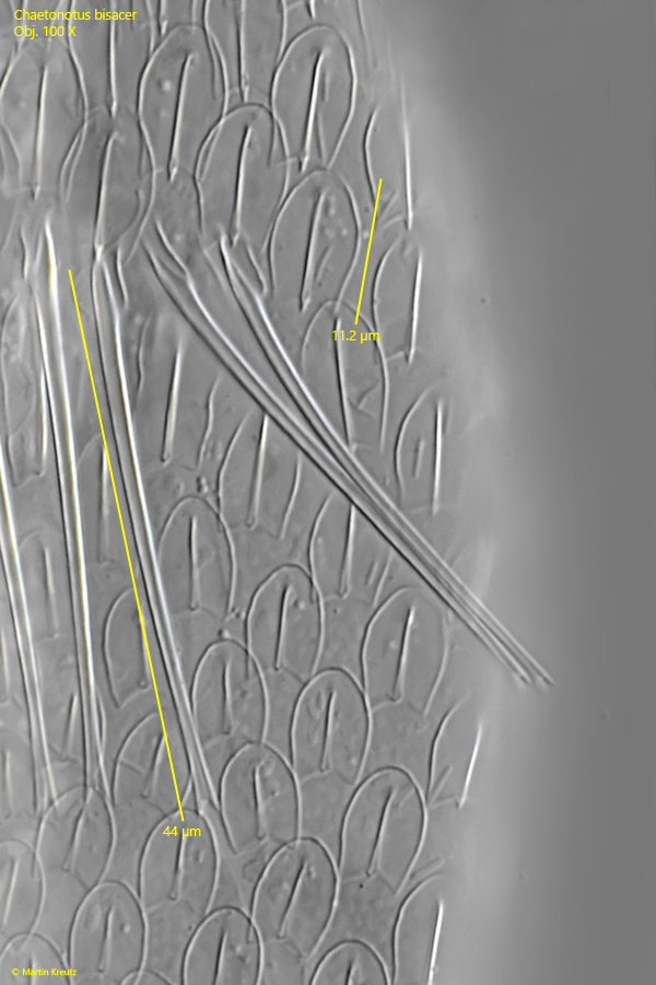
girdle spines 19–41 µm long
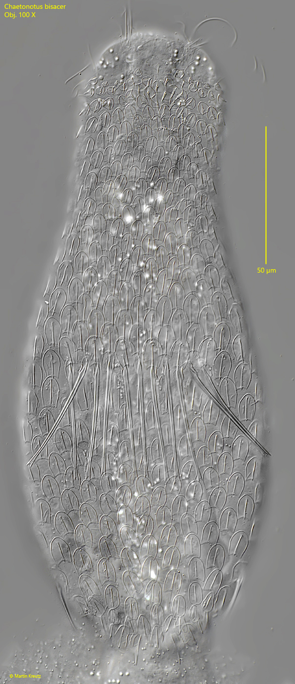
dorsal 11 longitudinal rows of keeled scales
dorsal scales slightly notched distally
dorsal anal region with field of elongated and triangular keeled scales
furca with wide straight notch
toes with short base (17–30 μm), long adhesive tubes
posterior 2 pairs of lateral, long spines
ventral 11–13 rows of elongated, narrow keeled scales
ventral terminal plates absent
Chaetonotus bisacer
Chaetonotus bisacer is described in the literature (Schwank, 1990) as a globally distributed gastrotrich, which has already been detected on various continents. However, the species seems to be extremely rare at my sampling sites. So far, I have only found 2 specimens in April 2026 in Ulmisried. There, the specimens were found in decayed sludge from decomposed leaves.
The most characteristic feature of Chaetonotus bisacer is a straight, transverse row of long spines, which runs approximately in the middle of the dorsal side. These spines are straight and have a double tip at the distal end. In my specimens, these spines were between 38–44 µm long (s. figs. 2, 6 and 7).
The scales on the dorsal side have a fairly homogeneous shape. They are shield-shaped, keeled, and have a notch at the distal end. In the middle of the body, they were about 11 µm long in my specimens (s. fig. 7). On the ventral side, there are narrow, keeled scales with a length of 5–6 µm (s. figs. 10 b and 11 b).
In freely swimming specimens, two long spines can be seen at the posterior end (s. figs. 10 a-b). These are the primary lateral spines, which extend far beyond the toes. Slightly above them is another pair of secondary, lateral spines, which are, however, much shorter (s. fig. 9). Between the toes, the body appears transversely truncated.
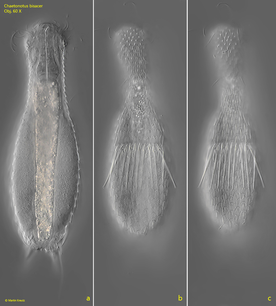
Fig. 1 a-c:Chaetonotus bisacer. L = 216 µm. Three focal planes of a freely swimming specimen from dorsal. Obj. 60 X.
Fig. 2:Chaetonotus bisacer. L = 216 µm. Dorsal view of the slightly squashed specimen as shown in fig. 1 a-c. Obj. 60 X.
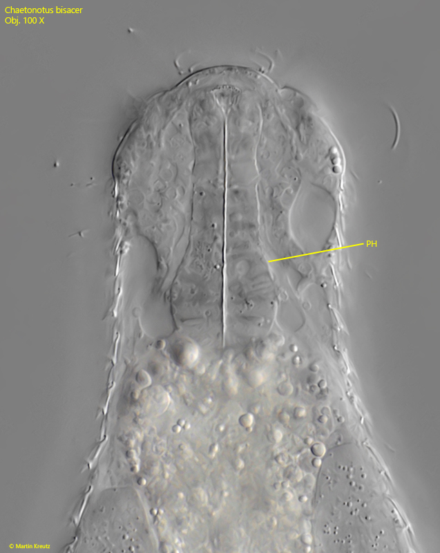
Fig. 3:Chaetonotus bisacer. The dumbbell-shaped pharynx (PH) of this specimen is 52 µm long. Obj. 100 X.
Fig. 4:Chaetonotus bisacer. Overall view of the dorsal scales of a squashed specimen. Obj. 100 X.
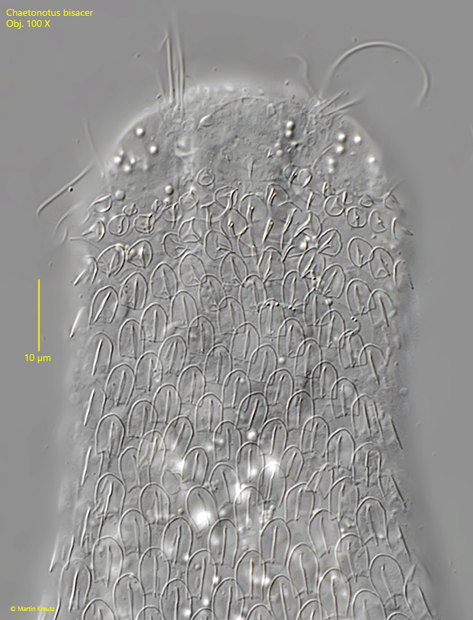
Fig. 5:Chaetonotus bisacer. The dorsal scales of the head- and neck-region in detail. Obj. 100 X.
Fig. 6:Chaetonotus bisacer. The transverse dorsal girdle in this specimen consists of 11 straight spines (1–11), which have a double tip at the distal end. Obj. 100 X.
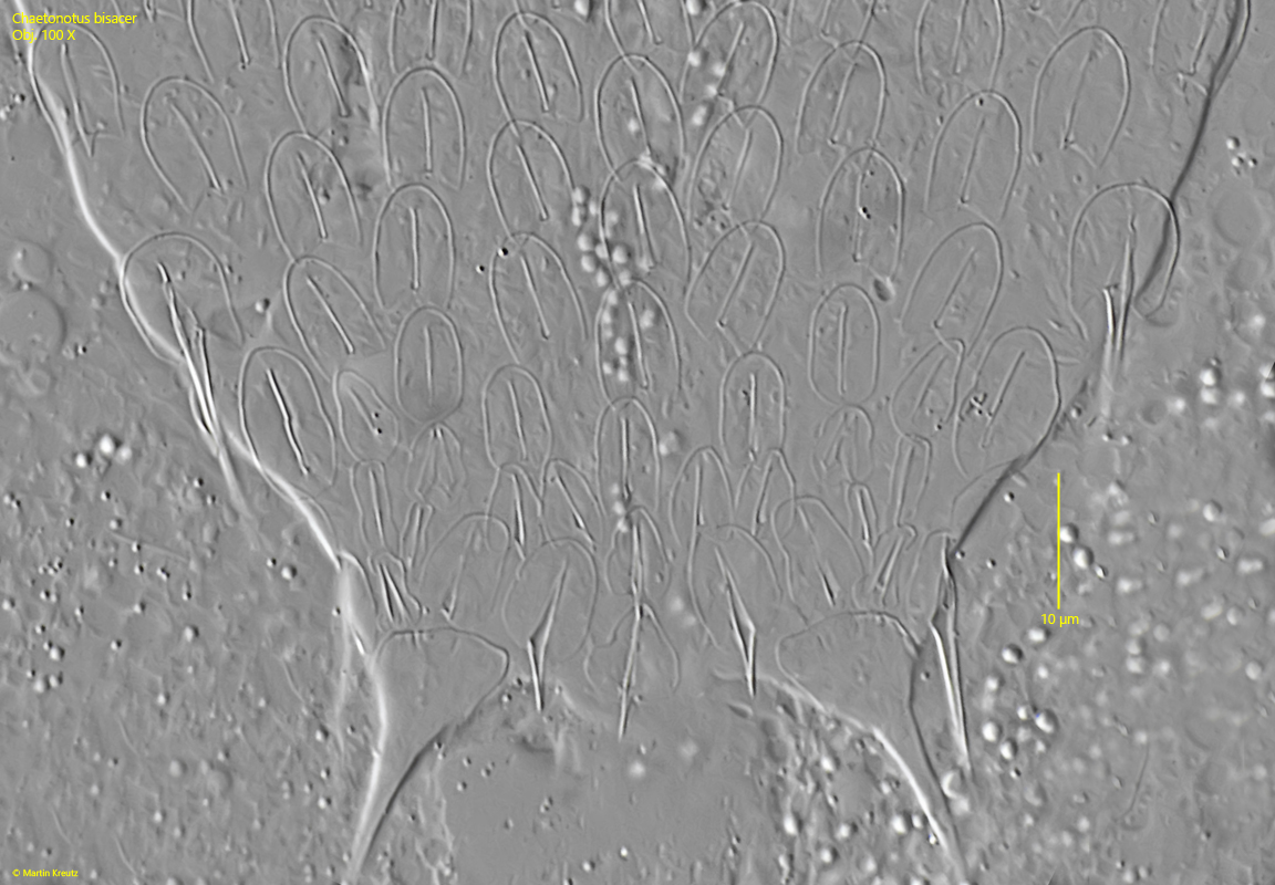
Fig. 7:Chaetonotus bisacer. The shape and length of the dorsal scales as well as of the dorsal spines in detail. Obj. 100 X.
Fig. 8:Chaetonotus bisacer. The pattern of the dorsal scales in the anal region in detail. Obj. 100 X.
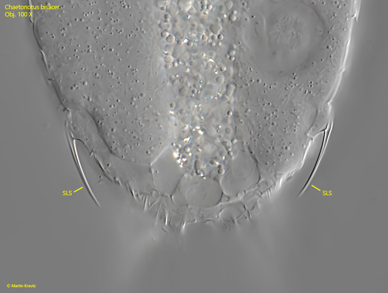
Fig. 9:Chaetonotus bisacer. The secondary lateral spines (SLS) near the posterior end. Obj. 100 X.
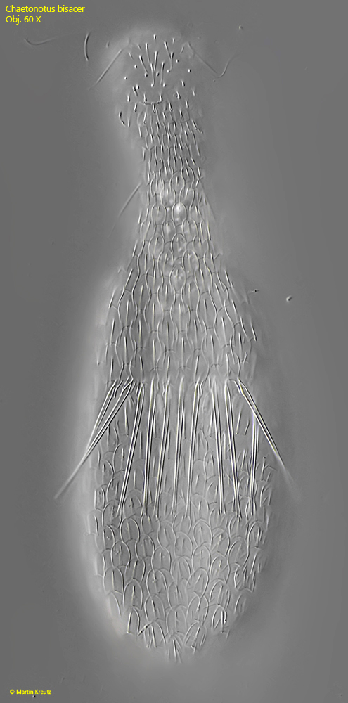
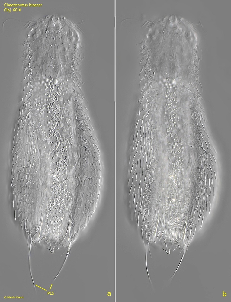
Fig. 10 a-b:Chaetonotus bisacer. L = 210 µm. Two focal planes of a slightly squashed specimen from ventral. Note the two long primary lateral spines (PLS) at the posterior end. Obj. 60 X.
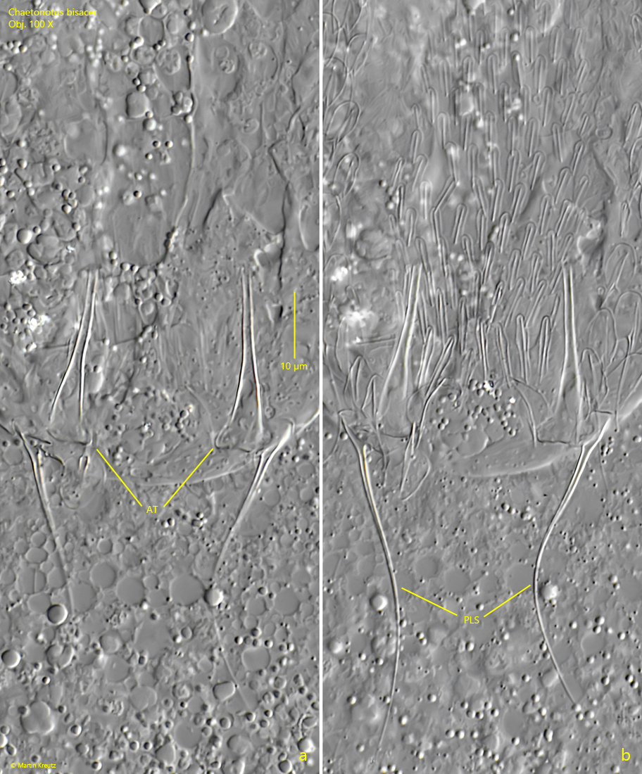
Fig. 11 a-b:Chaetonotus bisacer. Two focal planes of the scale pattern in the anal region on the ventral side. The scales in the anal region are narrow, keeled and about 5–6 µm long. AT = adhesive tubes (folded onto the ventral side), PLS = primary lateral spines. Obj. 100 X.