macroscopic thallus spherical, hemispherical or tubercular
thallus in a soft mucilaginous envelope
diameter of thallus up to several centimeter
filaments of cells arranged radially, trichotomously branched at distal end
end of filaments are tapered, end in a bluntly pointed cell
some terminal cells bearing multicellular setae lacking chloroplasts
width of cells 6–15 µm
one parietal chloroplast
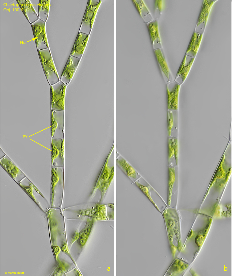
one to several pyrenoids
reproduction by quadriflagellate zoospores
Chaetophoropsis elegans
I find the macroscopically visible thalli of Chaetophoropsis elegans very frequently in the Simmelried. The thalli are either free-floating, lying loosely on the layer of mud, or attached to other aquatic plants and pieces of wood. Most of the time, I only find thalli that are a few millimeters in size, more or less spherical in shape.
The green alga Chaetophoropsis elegans was first described by Roth in 1806 as Chaetophora elegans. In 2019, it was renamed Chaetophoropsis elegans by B.W. Liu et al. based on genetic and morphological studies.
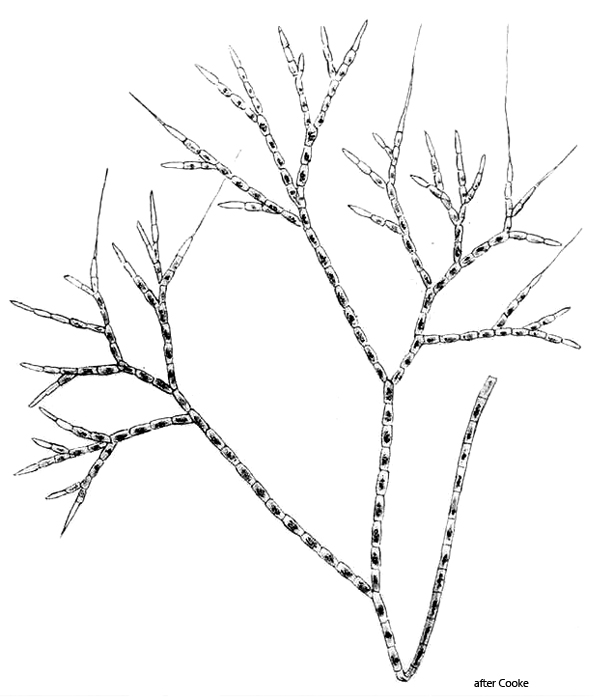
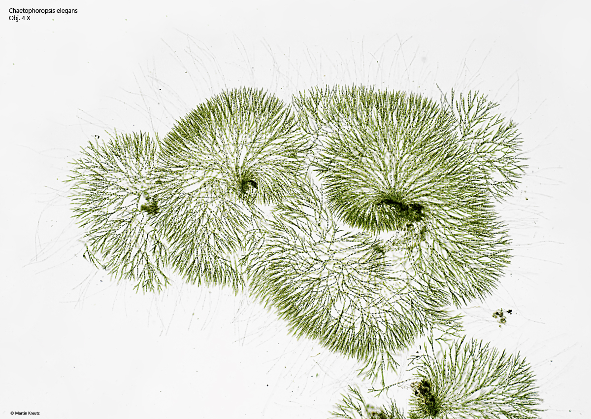
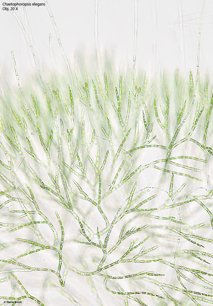
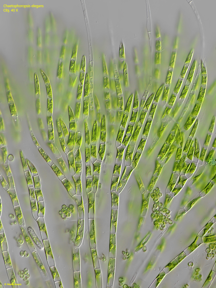
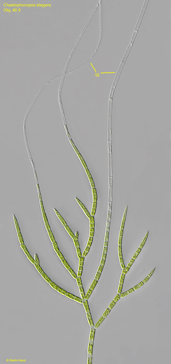
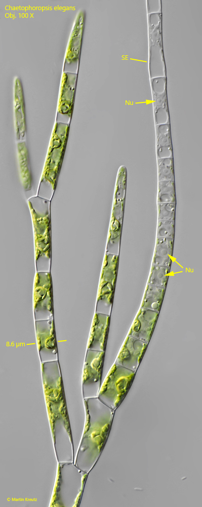
The thalli of Chaetophoropsis elegans consist of radially arranged filaments, which branch repeatedly towards the end. This results in a dense arrangement of branched filaments on the outside (s. figs. 2 and 3). The ends of these filaments taper to a terminal cell, which is bluntly pointed (s. fig. 3). However, some filaments do not end in terminal cells but instead form a seta (s. figs. 4 and 5). This is formed by repeated cell division, with the chloroplast of the cell being increasingly degraded after each division (s. fig. 5). In the distal cells of the seta, the protoplasts eventually die off as well, leaving behind empty, colorless cells that form the seta.
The thalli of Chaetophoropsis elegans are often colonized by unicellular algae, ciliates, and rotifers. Therefore, it is always worthwhile to examine the thalli more closely. In addition to ponds and bogs, Chaetophoropsis elegans is also native to freshwater aquariums, where it colonizes aquatic plants, wood, or glass as light green balls or cushions. It is undesirable among aquarists but difficult to remove.
Fig. 1:Chaetophoropsis elegans. D = 460–2100 µm (of thalli). Overview of three squashed thalli. Obj. 4 X.
Fig. 2:Chaetophoropsis elegans. A section of a thallus as shown in fig. 1. Obj. 20 X.
Fig. 3:Chaetophoropsis elegans. The branched distal ends of filaments. The terminal cells are tapered and bluntly pointed. Obj. 40 X.
Fig. 4:Chaetophoropsis elegans. A branched filament with some setae (SE) lacking chloroplasts. Obj. 20 X.
Fig. 5:Chaetophoropsis elegans. The formation of a seta (SE) from cells containing chloroplasts. As the seta grows, the chloroplasts are continuously degraded. With ongoing cell division, the protoplasts also die, so that at the distal end, empty cells form the setae. Nu = nucleus. Obj. 100 X.
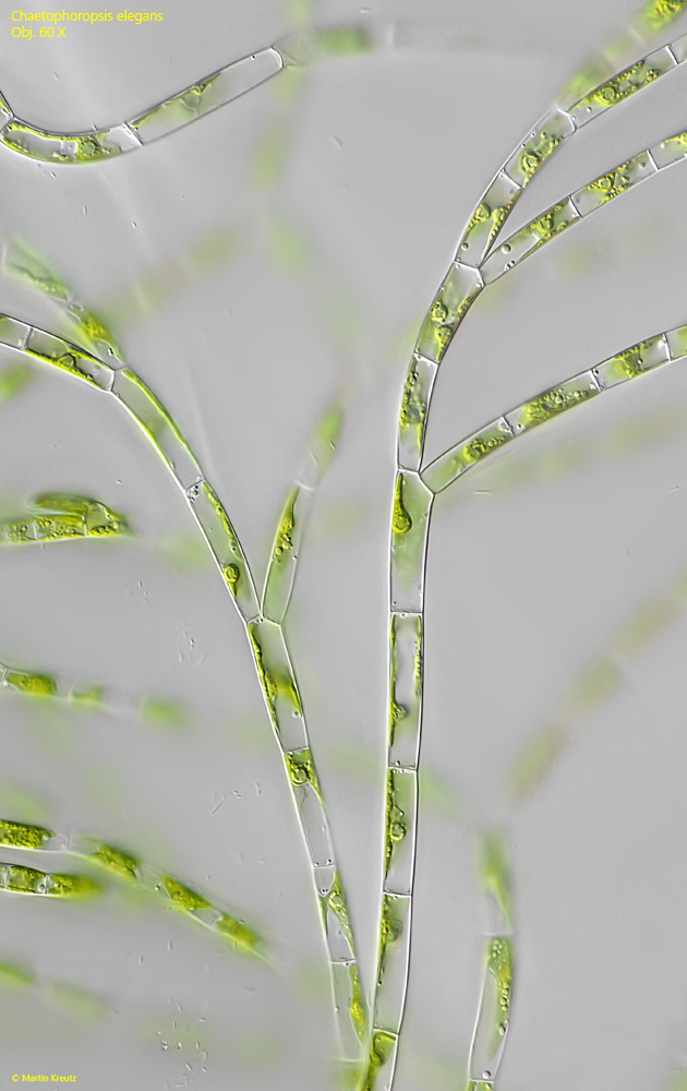
Fig. 6:Chaetophoropsis elegans. Two filaments near the center of the thallus. Obj. 60 X.
Fig. 7 a-b:Chaetophoropsis elegans. Two focal planes of a branched filament near the center of the thallus. Nu = nucleus, PY = pyrenoid. Obj. 100 X.
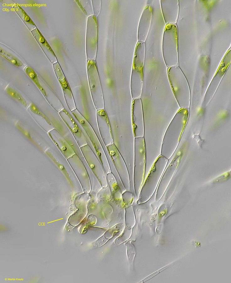
Fig. 8:Chaetophoropsis elegans. The colony center (CCE) with elongated cells serving as a rhizoid to fix the thallus to a substrate. Obj. 60 X.