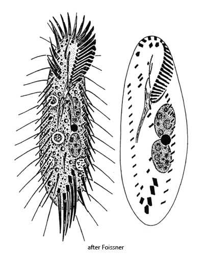
body long elliptical, slightly spindle-shaped, flexible
length 55–100 µm, width 15–30 µm
2 macronuclei, one micronucleus between them
contractile vacuole about mid-body, left margin
in anterior and posterior quarter often ring-shaped structure (“fat-body”)
left and right marginal rows
3 frontal cirri
4 frontoventral cirri
1 buccal cirrus
2 (sometimes 3) ventral cirri
3 postoral cirri
5 transversal cirri
Tachysoma pellionellum
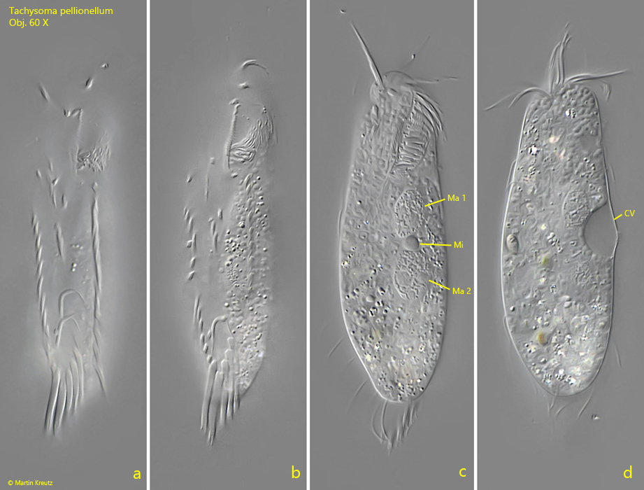
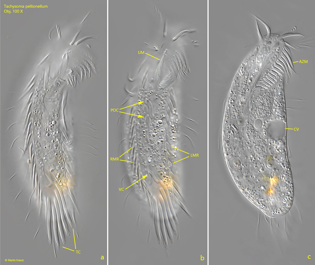
Taychsoma pellionellum is a very common hypotrichous ciliate that likes to settle on the floating coverslip and can then be observed very well. However, the arrangement of the cirri on the ventral side can only be seen at high magnification. Characteristic are a right and left marginal row. On the central ventral field there are only 3 postoral cirri and 2 ventral cirri, which are arranged just in front of the 5 transversal cirri (s. fig. 2 a-c). Two macronuclei are present, between which the round micronucleus is located (s. fig. 1 c).
Tachysoma pellionellum often shows a ring-shaped, conspicuous structure in the anterior or posterior quarter of the body. This is called the fat body. It is assumed that this is a storage body. However, I was not able to detect any fat bodies in my population of Tachysoma pellionellum.
The contractile vacuole of Tachysoma pellionellum is located almost in the middle of the body on the left side (s. figs. 1 d and 2 c), which is an important distinguishing feature from the similar species Holosticha pullaster. In this species the contractile vacuole lies clearly below the middle of the body. In addition, Holosticha pullasterhas 2 micronuclei instead of only one.
Fig. 1 a-d:Tachysoma pellionellum. L = 91 µm. Different focal planes of a freely swimming specimen from ventral. CV = contractile vacuole, Ma1 + Ma 2 = macronuclei, Mi = micronucleus. Obj. 60 X.
Fig. 2 a-c:Tachysoma pellionellum. L = 76 µm. A second specimen from ventral. Note the contractile vacuole in mid-body at the left margin and the two ventral cirri (VC) above the 5 transversal cirri (TC). AZM = adoral zone of membranelles, LMR = left marginal row, POC = postoral cirri, RMR = right marginal row, UM = undulating membrane. Obj. 100 X.