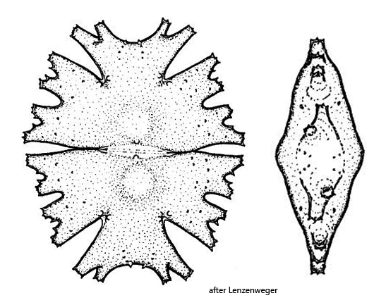
apical lobes are trapezoidal, set off from the lateral lobes
either side of the isthmus a distinct wart
pair of warts at base of notches to apical lobes
cell wall punctate
chloroplasts with scattered pyrenoids
Micrasterias americana
In June 2026, a mass development of Micrasterias americana was found by Michael Butkay in the Vogelsberger Weiher in Austria. He kindly made a subset of the sample available to me. In the samplings sites in my area I have not yet been able to find Micrasterias americana.
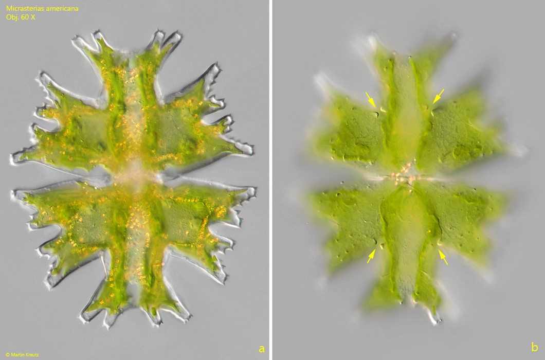
Micrasterias americana can easily be recognized by the clearly separated apical lobes, which are distinctly set apart from the lateral lobes by a neck-shaped extension. The ends of the lobes bear only short spines, and the indentations between the secondary lobes are approximately U-shaped. In addition, at the base of the indentations leading to the apical lobes there is in each case a pair of warts, which can be clearly seen when the focal planes is adjusted on the cell surface (s. fig. 1 b).
Fig. 1 a-b:Micrasterias americana. L = 150 µm. Two focal planes of a specimen in DIC. Note the pair of warts at the base of the notches of the apical lobes (arrows). Obj. 60 X.
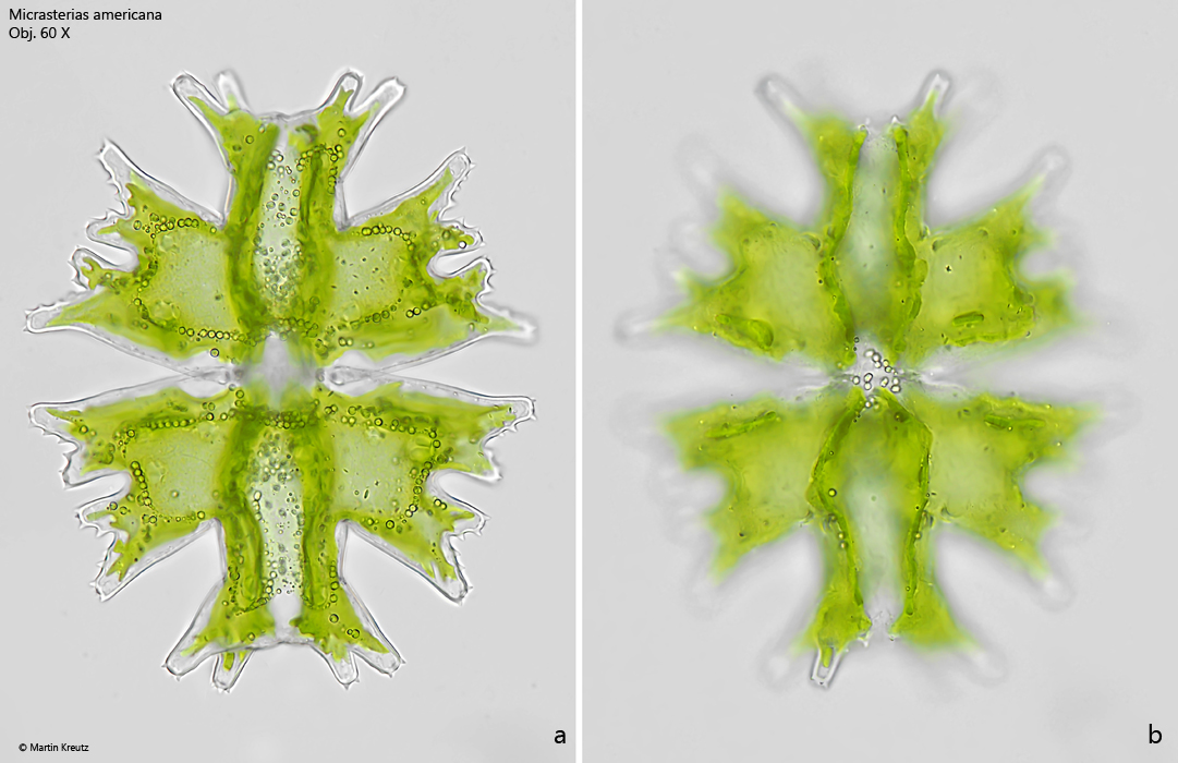
Fig. 2 a-b:Micrasterias americana. L = 150 µm. The same specimen as shown in fig. 1 a-b in brightfield illumination. Obj. 60 X.