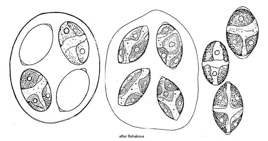
cells ellipsoidal to broad spindle-shaped, sometimes with thickened apices
apices sharply rounded
cell in gelatinized and expanded mother cell wall
length 6.4–32 µm, width 4–22 µm.
2–4 parietal chloroplasts, each with one pyrenoid
older cells with 8 chloroplasts (before cell division)
Oocystis marssonii
So far I have only been able to find Oocystis marssonii in the Botanical Garden Darmstadt, although the species is described as common and widespread. At the sampling sites in my local area, I have so far been unable to detect Oocystis marssonii.
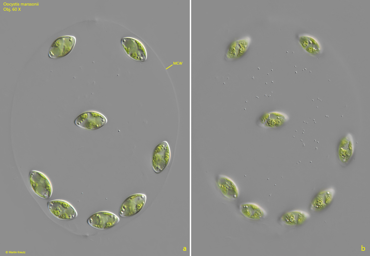
The cells of Oocystis marssonii are broadly spindle-shaped, usually with slightly thickened apices. The cell wall of the mother cell gelatinizes during cell division, and the daughter cells are then located in the swollen envelope. Depending on the number of divisions, 2, 4, or 8 cells may be present in it. In my population, I was only able to find colonies with 8 cells.
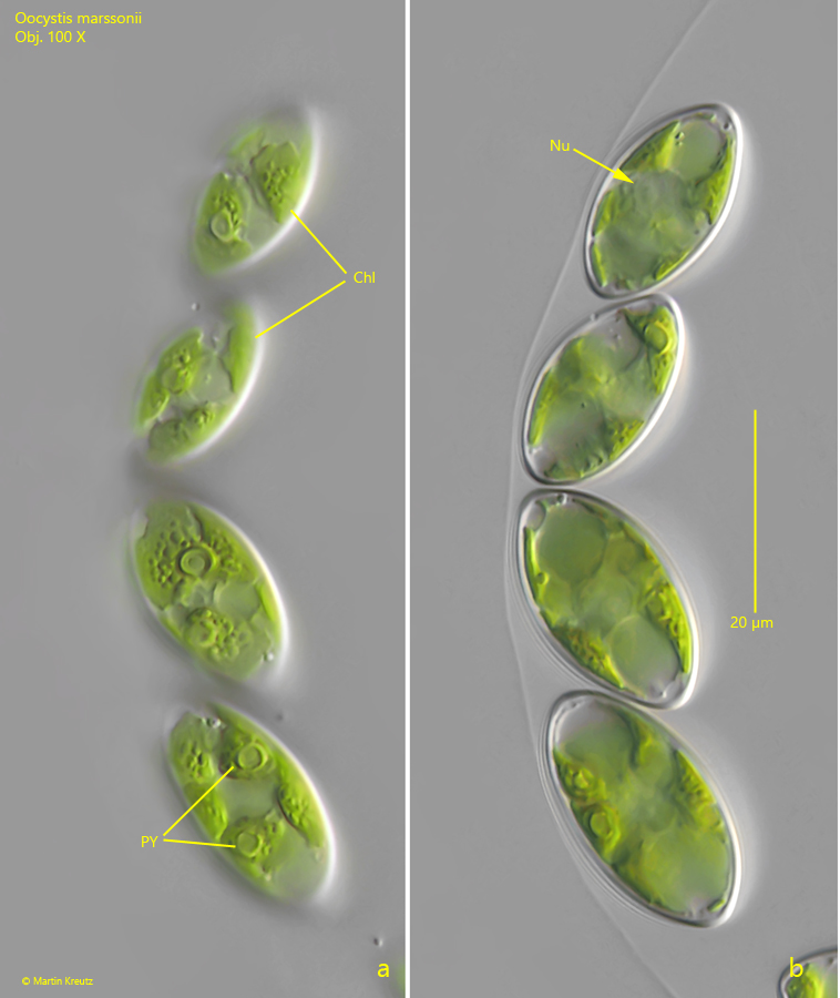
An essential characteristic of Oocystis marssonii is the size of the cells, which in my population was between 20–26 µm. Each cell contains 2–4 chloroplasts with slightly wavy margins that lie against the cell wall (parietal). Each chloroplast contains a clearly visible pyrenoid.
The likewise widespread species Oocystis lacustris has substantially smaller cells, at 6–11 µm in length, and only one chloroplast with a pyrenoid (before division 3–4 chloroplasts).
Fig. 1 a-b:Oocystis marssonii. L = 160 µm (of colony). A colony of 8 cells enclosed in the gelitinized cell wall of the mother cell (MCW). Obj. 60 X.
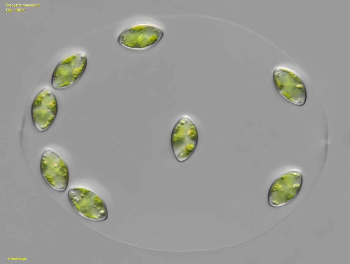
Fig. 2:Oocystis marssonii. L = 160 µm (of colony). The colony as shown in fig. 1 a-b at higher magnification. Obj. 100 X.
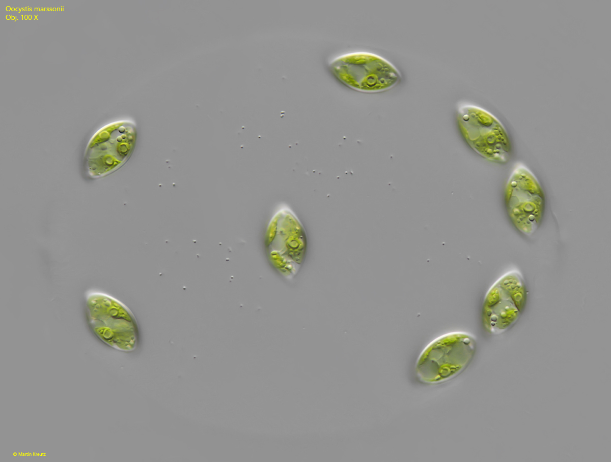
Fig. 3:Oocystis marssonii. L = 160 µm (of colony). The colony as shown in fig. 1 a-b at higher magnification. Obj. 100 X.
Fig. 4 a-b:Oocystis marssonii. L = 22-26 µm (of cells). Some cells of a second colony in detail. In the cells 3–4 chloroplasts are visible each with a pyrenoids (PY). Nu = nucleus. Obj. 100 X.