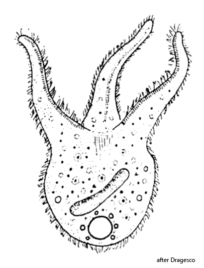
body campanulate with three oral lobes (proboscides or tentacles)
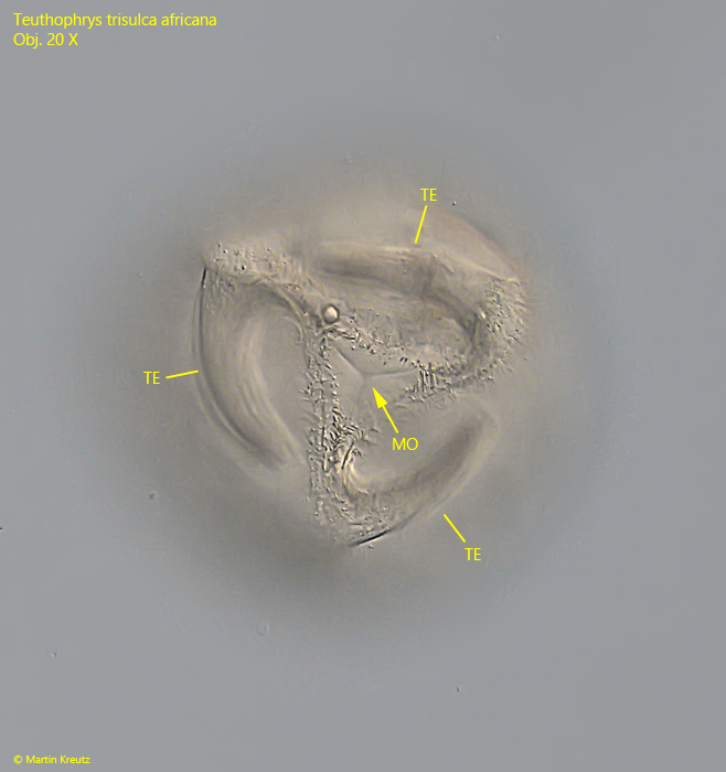
tentacles curved counterclockwise (in apical view)
length 150–300 µm
mouth opening in center between tentacles
macronucleus rope-shaped or slightly moniliform
contractile vacuole terminal
symbiotic algae absent
extrusomes rod-shaped, about 6 X 0.8 µm
dorsal brush with 3 rows of bristles (need silver impregnation)
Teuthophrys trisulca africana
The specimens of Teuthophrys trisulcata africana shown below come from a culture by Holger Weis, who kindly made some specimens available to me. The basis of his culture is samples from Anne Gleich from a pond near Mackenbach, close to Kaiserslautern. In the plankton of this pond, Teuthophrys trisulca africana as well as Teuthophrys trisulca trisulca were found in the months of March to June in 2025 and 2026. Both species occurred in the pond at the same time. The appearance of the Teuthophrys populations coincided with the increase of populations of various rotifer species, the preferred prey of this ciliate. I myself have so far been unable to find either Teuthophrys trisulca africana or Teuthophrys trisulca trisulca.
Most populations of Teuthophrys trisulca detected so far have contained symbiotic algae. It was not until 1972 that Dragesco described a population without symbiotic algae. Foissner (1999) demonstrated that populations of Teuthophrys trisulca with symbiotic algae form cysts which contain the symbiotic algae and thus pass them on to the next generation. From this he concluded that the presence or absence of symbiotic algae is a permanent characteristic in the populations. As a consequence, he named the populations with symbiotic algae Teuthophrys trisulca trisulca and those without symbiotic algae Teuthophrys trisulca africana.
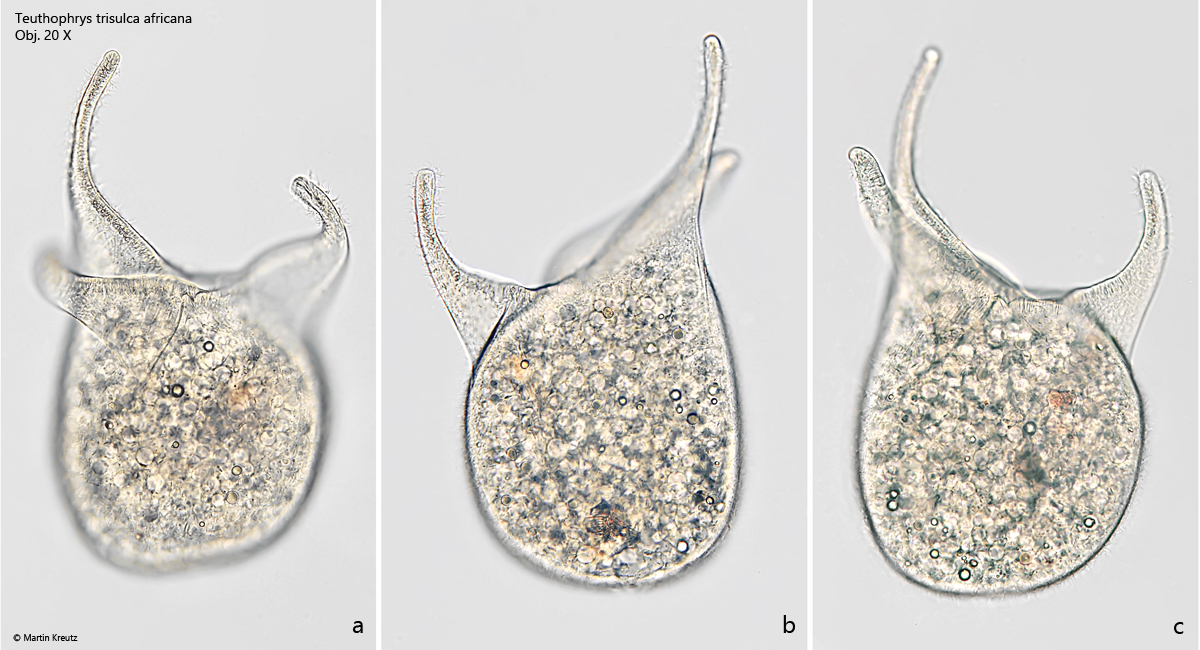
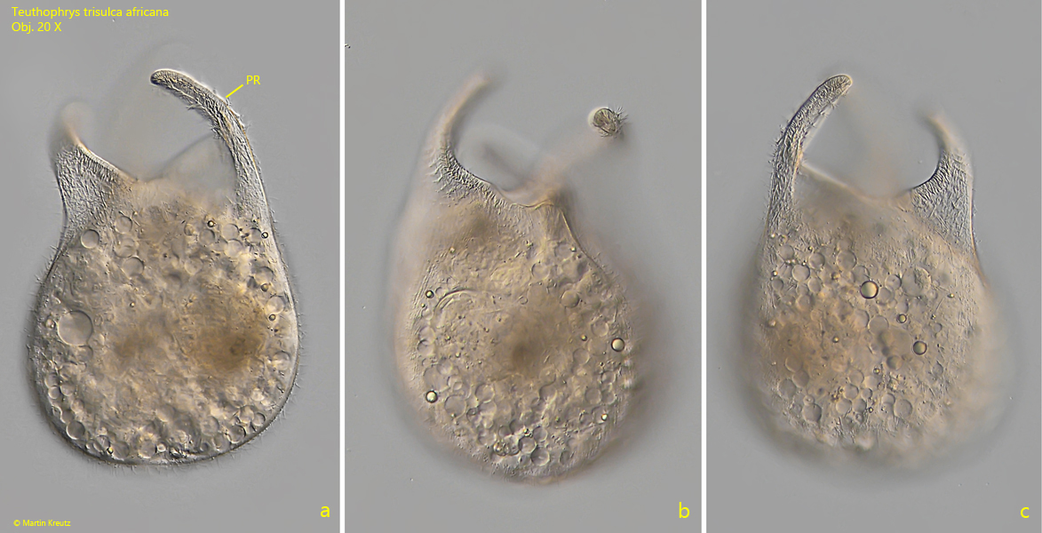
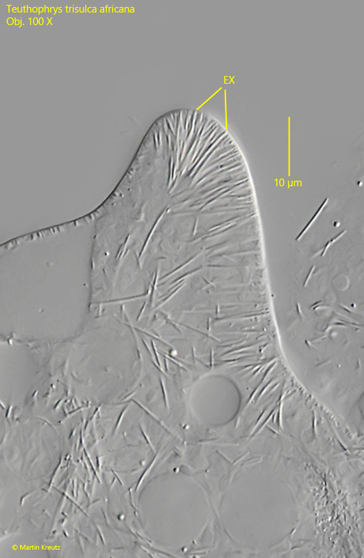
The specimens I examined did not contain symbiotic algae, which means they must be Teuthophrys trisulca africana. Confusion with other species is excluded because Teuthophrys has three apical “tentacles” as a unique distinguishing feature (s. fig. 1 a-c). The posterior part of the body is approximately sac-shaped. Each tentacle is provided with a fringe of extrusomes (s. fig. 7). The mouth opening is located as a triangular slit in the middle between the tentacles (s. fig. 3). The natural body shape of Teuthophrys trisulca africana is difficult to document, since placing a coverslip on it leads to immediate contraction of the body or even to bursting. Therefore, the images shown in fig. 1 a–c and fig. 2 a–c were taken of freely swimming specimens without a coverslip.
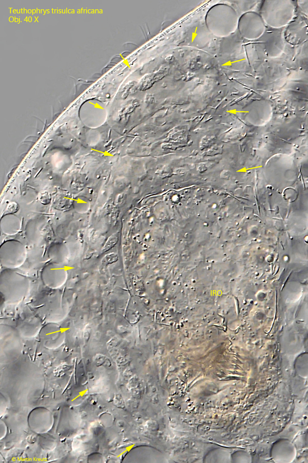
Because of the sensitivity to the coverslip, I was only able to document the shape of the macronucleus inadequately. It begins to deform very quickly and eventually breaks up into several parts. However, I was able to determine that it is about 200–240 µm long and rope-shaped. This shape and size are supported by the videos by Ariel Qassis (s. below), in which the macronucleus can be clearly seen. Dragesco (1972) describes an African population with a dumbbell-shaped macronucleus. The shape of the macronucleus therefore seems to be variable. I was not able to detect the micronucleus (or micronuclei). Earlier authors have not described them either.
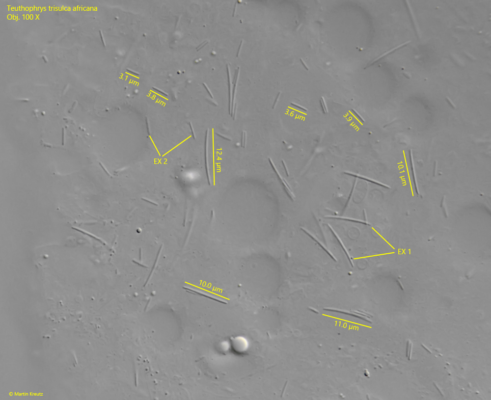
Deviating from Foissner’s description of Teuthophrys trisulca africana, I was able to distinguish two types of extrusomes. Type 1 consists of very slightly curved rods with a length of 10–13 µm, while type 2 consists of straight, short rods with a length of 3–4 µm (s. fig. 9). Foissner mentions only rods with a length of 6 µm.
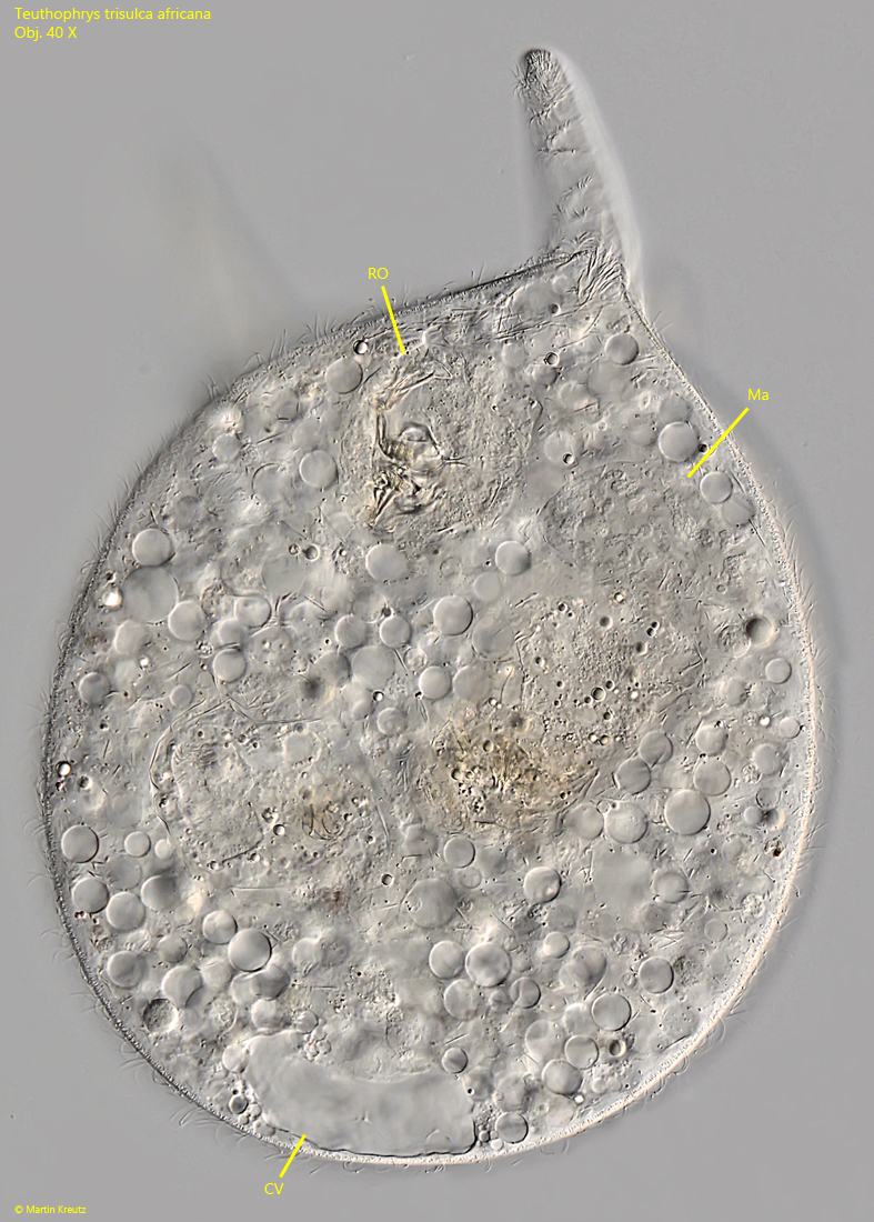
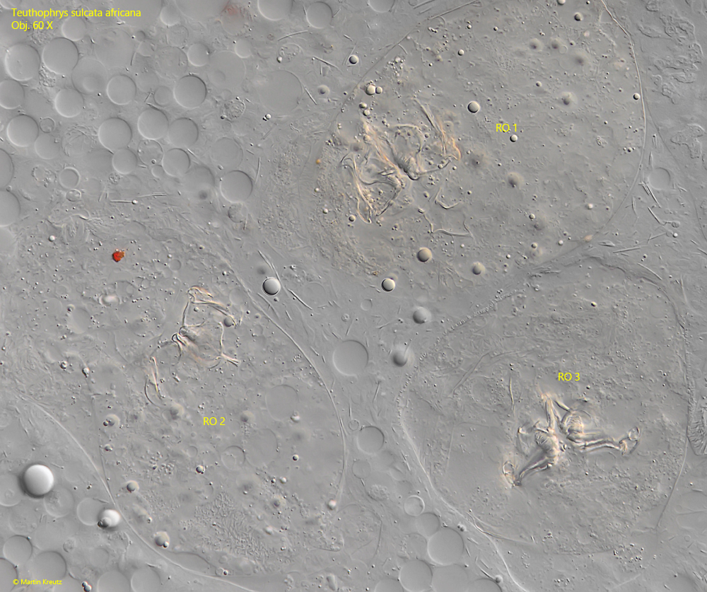
In the cytoplasm one finds very many ingested rotifers, the main food source of Teuthophrys trisulca africana. They can be easily recognized by the indigestible trophi. These consist mainly of Brachionus spec. and Keratella spec., which served as food organisms for the culture.
Several videos of Teuthophrys trisulca africana were recorded by Ariel Qassis:
Fig. 1 a-c:Teuthophrys trisulca africana. L = 386 µm. A freely swimming specimen with erected tentacle in brightfield illumination. Obj. 20 X.
Fig. 2 a-c:Teuthophrys trisulca africana. L = 298 µm. A second freely swimming specimen in DIC. Obj. 20 X.
Fig. 3:Teuthophrys trisulca africana. The same specimen as shown in fig. 2 a-c in apical view. The mouth opening (MO) is a trigonal slit in the center of the tentacles (TE). Note that the tentacles are curved counterclockwise. Obj. 20 X.
Fig. 4:Teuthophrys trisulca africana. A slightly squashed specimen. The contractile vacuole (CV ) is located terminal. In the cytoplasm the remains of ingested rotifers (RO) are visible. Ma = macronucleus. Obj. 40 X.
Fig. 5:Teuthophrys trisulca africana. The deformed macronucleus (arrows) is rope-shaped and about 240 µm long. Obj. 40 X.
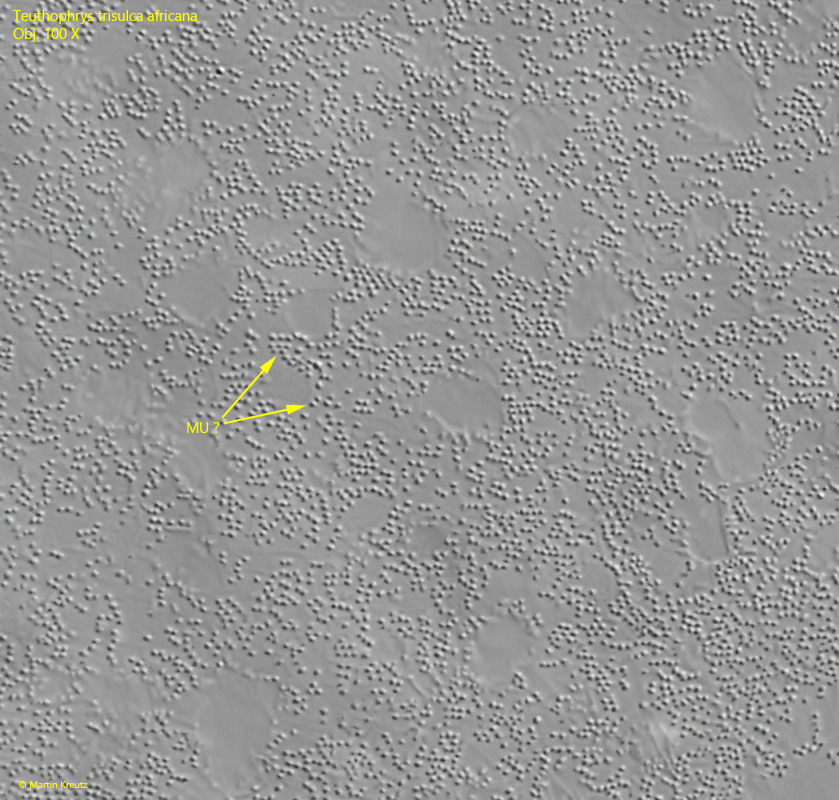
Fig. 6:Teuthophrys trisulca africana. Focal plane on the cortex of a squashed specimen. Numerous granules are visible with a diameter of about 1 µm, likely mucocysts (MU ?). Obj. 100 X.
Fig. 7:Teuthophrys trisulca africana. Three ingested rotifers in the cytoplasm of a squashed specimen. In each rotifer the indigestible trophi can be recognized. Obj. 60 X.
Fig. 8:Teuthophrys trisulca africana. A squashed tentacle with a fringe of extrusomes (EX). Obj. 100 X.
Fig. 9:Teuthophrys trisulca africana. In a strongly squashed specimen two types of extrusomes (EX 1, EX 2) are visible. The extrusomes of the type 1 (EX 1) are very slightly curved rods with a length of 10–13 µm long, while the extrusomes of the type 2 (EX 2) are straight rods with a length of 3–4 µm. Obj. 100 X.