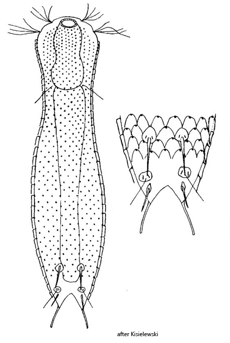
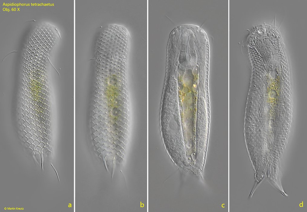
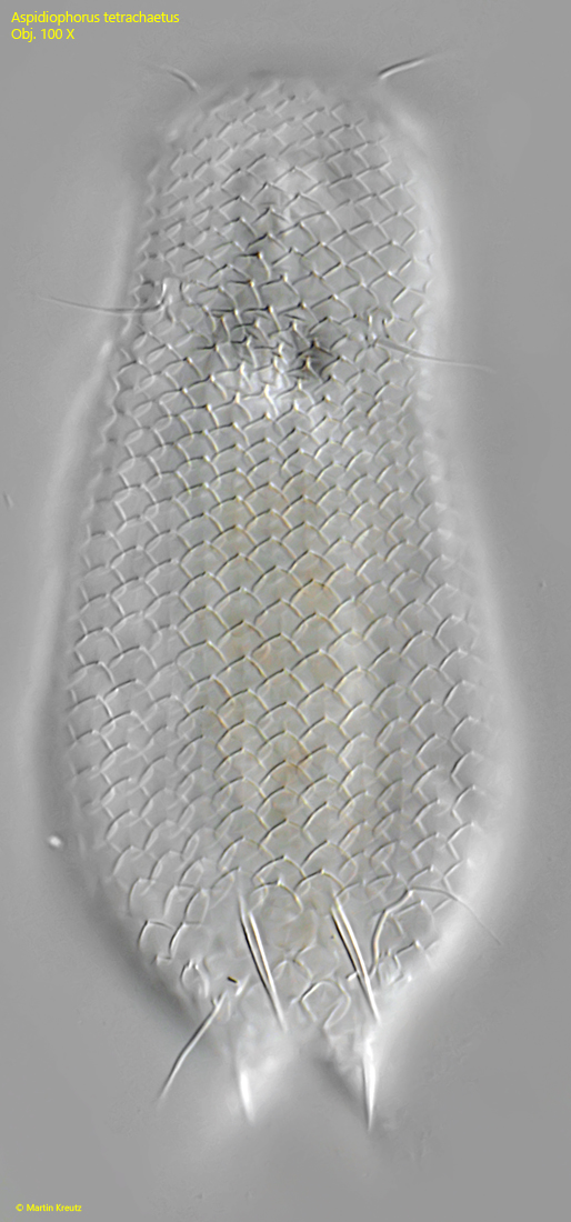
dorsal 15–17 longitudinal rows of diamond-shaped scales
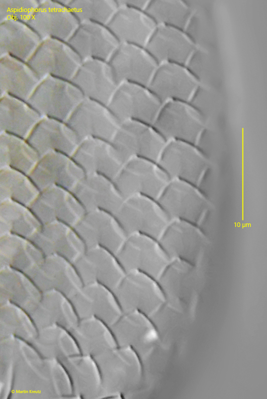
dorsal scales about 5 µm long
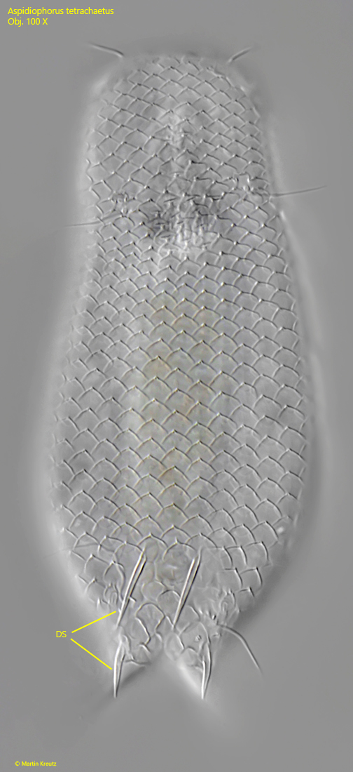
two pairs of dorsal spines near posterior end
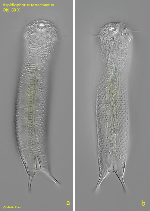
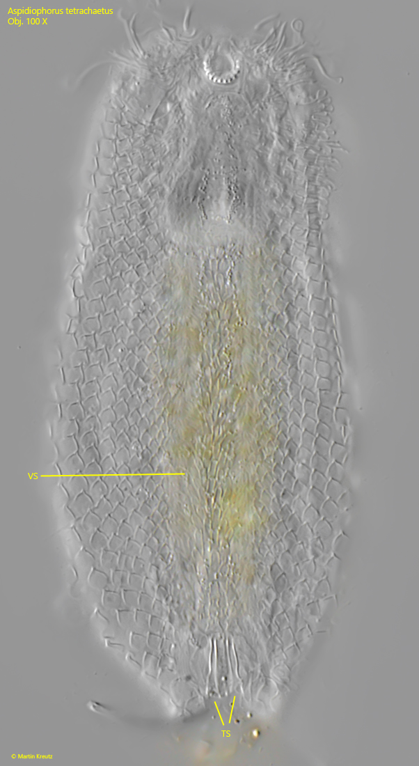
ventral field with 8–9 of very small scales (1.5–2.5 µm)
two ventral terminal scales
Aspidiophorus tetrachaetus
So far I have only been able to find Aspidiophorus tetrachaetus once, in a sample from the Schwemm Moor in June 2026. Among several samples from the Schwemm Moor, the specimens were present in only one of these samples. This contained only a thin sediment layer, consisting mainly of algae, cyanobacteria and bacteria.
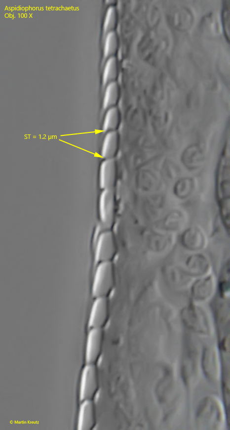
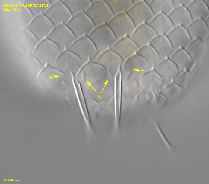
The gastrotrichs of the genus Aspidiophorus can be easily recognized by the stalked scales (s. fig. 7). Due to the stalks, the actual scales stand 1–2 µm away from the body. The species Aspidiophorus tetrachaetus is easily identifiable by 4 spines located on the dorsal side at the posterior end (s. fig. 8). This makes the species unmistakable. These spines sit on 4 oval scales that are keeled at their margins (s. fig. 9).
The stalked dorsal scales are diamond-shaped with rounded corners (s. fig. 6). They overlap each other and do not form any gap. According to my measurements, they are about 5 µm long, which corresponds well with the data given by Schwank (1990). On the ventral side, there are several parallel rows of scales about 3 µm long, whose exact shape is difficult to discern (s. fig. 10). Based on my observations, they appear to be oblong with parallel sides and a delicate keel. At the posterior end on the ventral side, there are 2 terminal scales with a distinct keel and lobe-shaped protrusions (s. fig. 10).
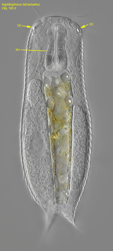
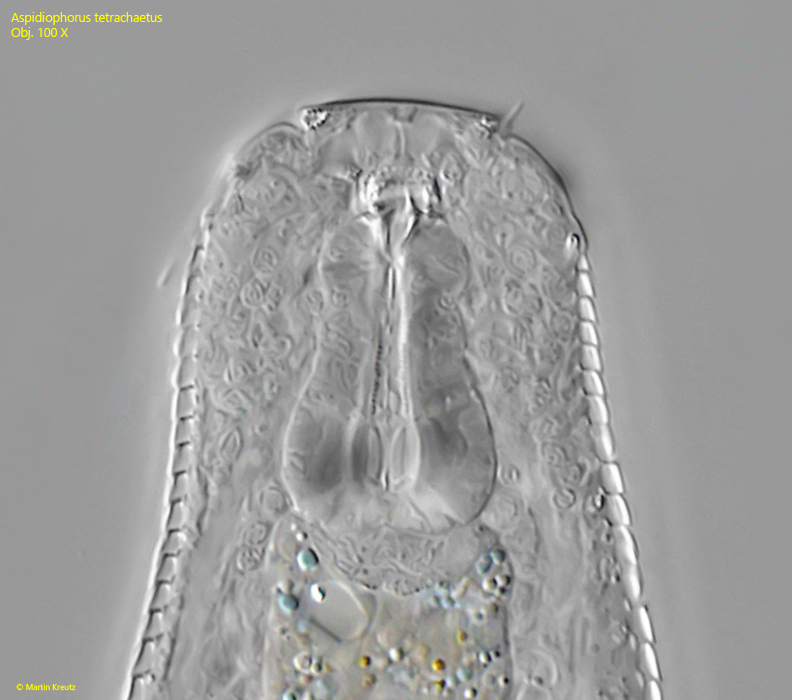
In deviation from the descriptions by Schwank (1990) and Kisielewski (2011), I was able to identify distinct ocelli at the anterior margin of the head in the specimens of my population, which were not mentioned by these authors. The ocelli had a granular character. All other characteristics correspond to the descriptions of Aspidiophorus tetrachaetus.
Fig. 1 a-d:Aspidiophorus tetrachaetus. L = 134 µm. A freely swimming specimen from dorsal. Obj. 60 X.
Fig. 2 a-b:Aspidiophorus tetrachaetus. L = 130 µm. A second freely swimming specimen from ventral. Obj. 60 X.
Fig. 3:Aspidiophorus tetrachaetus. L = 131 µm. Focal plane on the two ocelli (OC) at the front of the head. PH = pharynx. Obj. 100 X.
Fig. 4:Aspidiophorus tetrachaetus. The slightly squashed pharynx. Obj. 100 X.
Fig. 5:Aspidiophorus tetrachaetus. The dorsal scales of a slightly squashed specimen. Obj. 100 X.
Fig. 6:Aspidiophorus tetrachaetus. The dorsal scales are diamond-shaped with rounded edges. The scales are about 5 µm long. Obj. 100 X.
Fig. 7:Aspidiophorus tetrachaetus. The dorsal scales sit on short stalks (ST) with a length of 1.2 µm. Obj. 100 X.
Fig. 8:Aspidiophorus tetrachaetus. The same specimen as shown in fig. 6 with focal plane on the 4 dorsal spines (DS). The upper spines are 14 µm long while the lower spines are 9 µm long. Obj. 100 X.
Fig. 9:Aspidiophorus tetrachaetus. The upper dorsal spines arise from two oval scales (arrows) with each two small keels (KE). Obj. 100 X.
Fig. 10:Aspidiophorus tetrachaetus. The ventral scales are very small (about 3 µm) and hard to see. They seems to be oblong with parallel sides and an indistinct keel. At the posterior end two terminal scales (TS) are visible. Obj. 100 X.