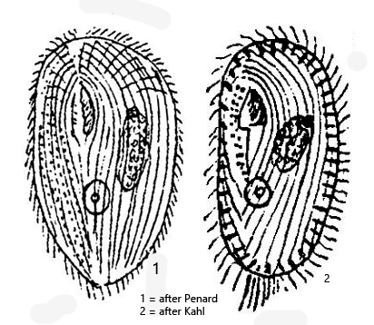
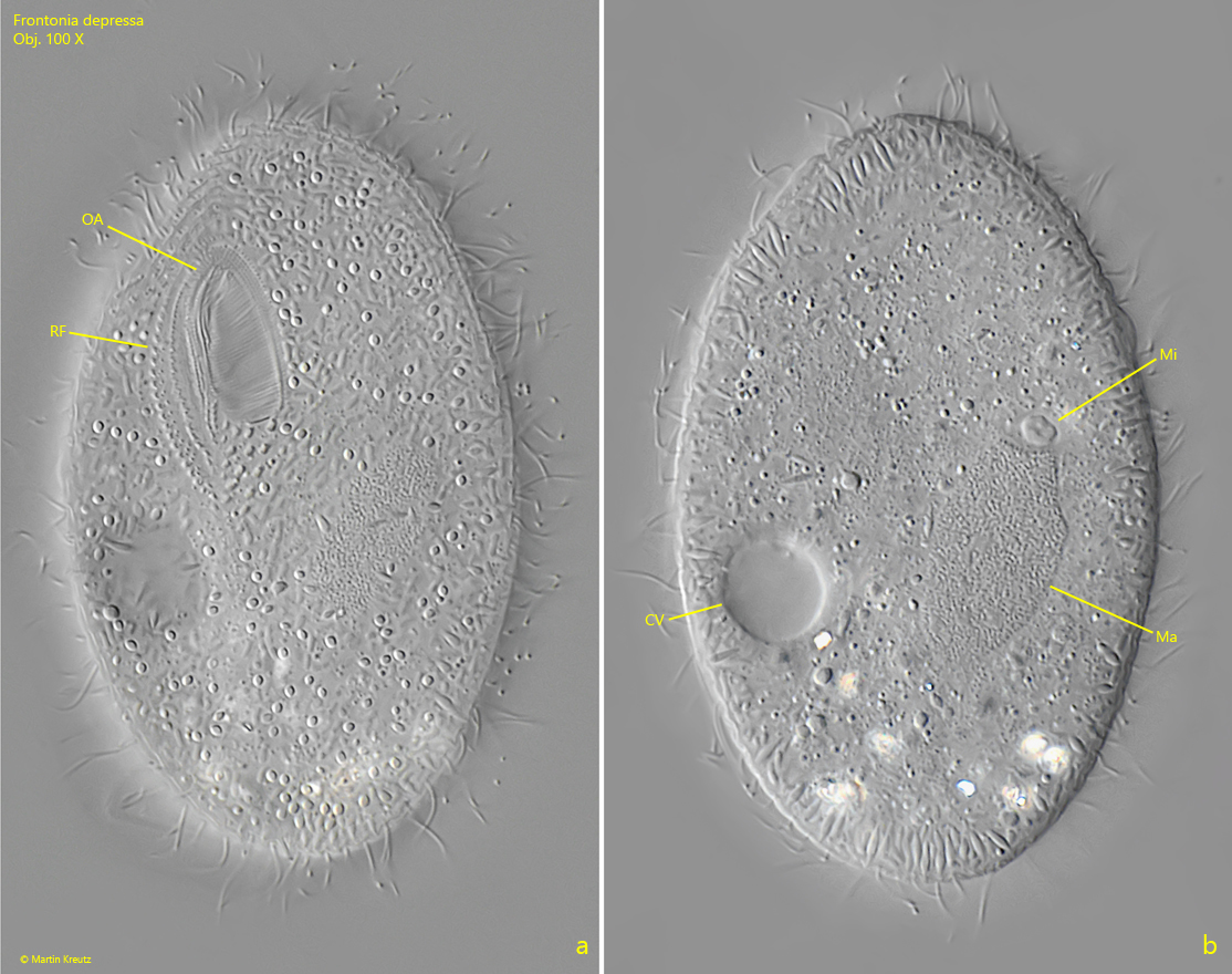
three distinct rows of fibrils right from mouth opening
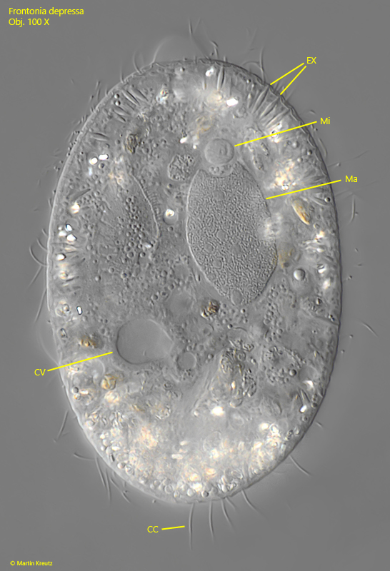
one contractile vacuole below cell equator
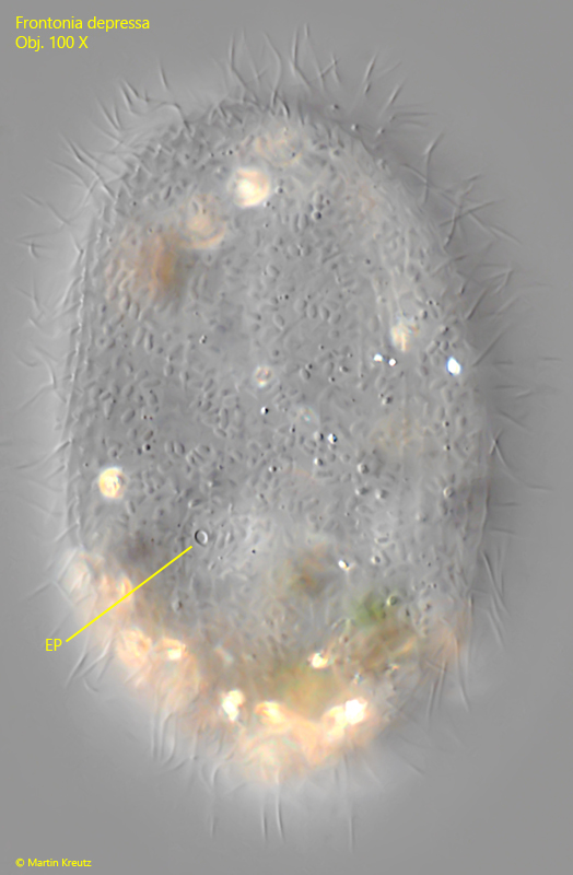
contractile vacuole with one excretion pore
macronucleus ellipsoid with slightly tapred ends
one micronuleus at end of macronucleus
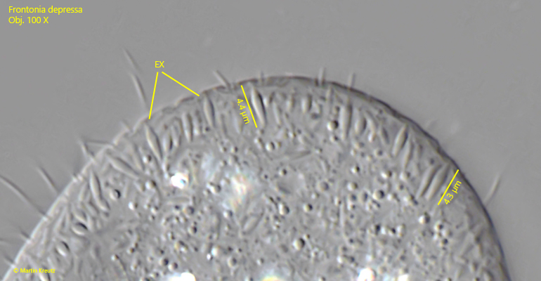
fringe of extrusomes beneath pellicle
several elongated caudal cilia
habitat moss
Frontonia depressa
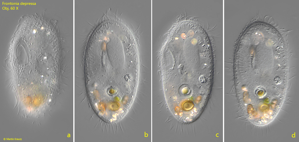
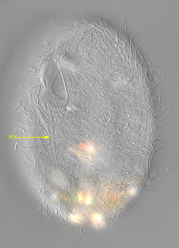
I found Frontona depressa in moss samples from trees in the years 2021, 2024 and 2025. This is the typical habitat of this ciliate, just as Kahl (1931) also described it. A typical characteristic, in addition to its small size and habitat, is the position of the contractile vacuole, which lies below the cell equator. It has only one excretory pore (s. fig. 7). At the posterior end there are some elongated cilia (s. fig. 6), which can also be clearly seen in free-swimming specimens (s. fig. 1 b). The preoral and postoral sutures (s. fig. 5) are only weakly developed in Frontonia depressa. I was only able to discern them faintly. According to my measurements, the spindle-shaped extrusomes are 4.2–4.9 µm long (s. fig. 8).
Most specimens in my populations were colorless and transparent, without food vacuoles. A few specimens had filled food vacuoles with spores of plants or fungi (s. fig. 1 a-d). Sometimes storage crystals could also be seen in the posterior third of the cell (s. fig. 6).
Fig. 1 a-d:Frontonia depressa. L = 67 µm. A freely swimming specimen from ventral. Obj. 60 X.
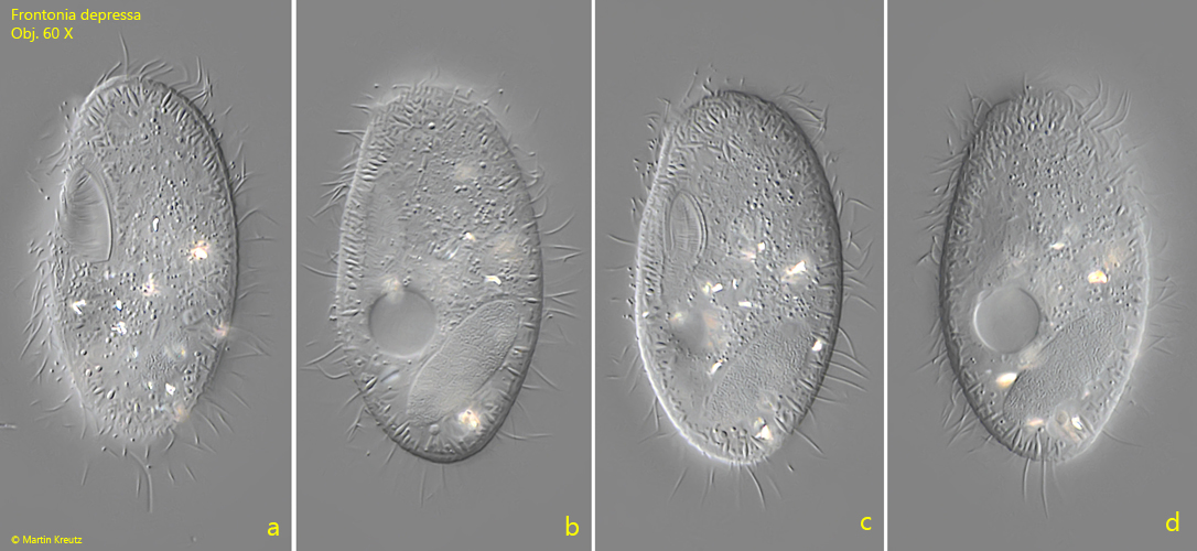
Fig. 2 a-d:Frontonia depressa. L = 71 µm. A second freely swimming specimen from ventral. Obj. 60 X.
Fig. 3 a-b:Frontonia depressa. Two focal planes of a slightly squashed specimen from ventral. Note the distinct rows of fibrils (RF) to the right of the oral apparatus (OA). The contractile vacuole (CV) is located below the cell equator. Ma = macronucleus, Mi = micronucleus. Obj. 100 X.
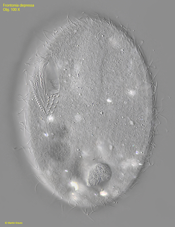
Fig. 4:Frontonia depressa. The oral apparatus in detail in a squashed specimen. Obj. 100 X.
Fig. 5:Frontonia depressa. Focal plane on the postoral suture (POS) in a slightly squashed specimen. The suture seems to extend only to the posterior third of the body. Obj. 100 X.
Fig. 6:Frontonia depressa. Focal plane on the macronucleus (Ma) and the micronucleus (Mi) in a squashes specimen. CC = caudal cilia, CV = contractile vacuole, EX = extrusomes. Obj. 100 X.
Fig. 7:Frontonia depressa. The single excretion pore (EP) of the contractile vacuole is located dorsal, observed from ventral. Obj. 100 X.
Fig. 8:Frontonia depressa. The extrusomes (EX) are spindle shaped with a length of 4.2–4.9 µm. Obj. 100 X.