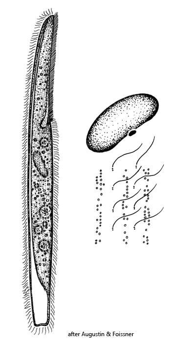
contractile vacuole terminal with a dorsal collecting canal
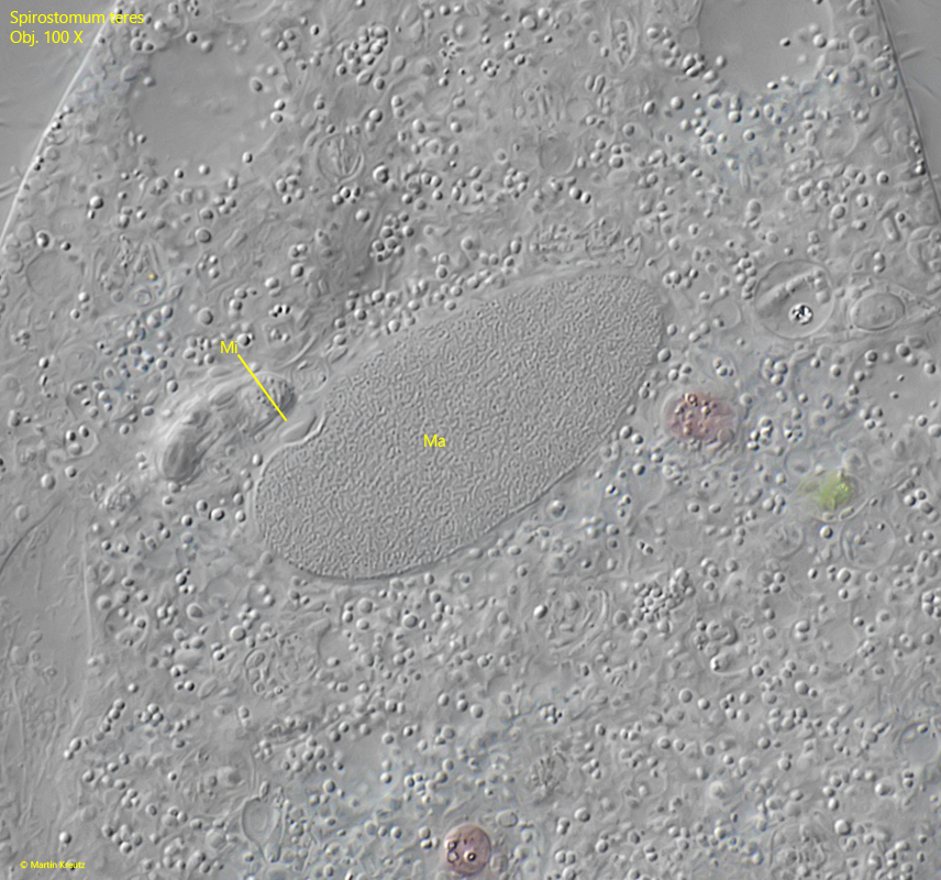
macronucleus ellipsoid
1–2 lens-shaped micronuclei
25–30 longitudinal rows of cilia
rows of yellowish cortical granules between rows of cilia
cortical granules in scattered within rows
oral groove about 40–50 % of body length
adoral zone on left side of oral groove
Spirostomum teres
I find Spirostomum teres mainly in habitats where a thick layer of fallen leaves is present on the bottom. The specimens occur most frequently in this decomposing mass of leaves. Compared to Spirostomum minus and Spirostomum ambiguum, however, Spirostomum teres is considerably rarer in my habitats.
Spirostomum teres possesses an ellipsoidal macronucleus (s. fig. 6) and can therefore be easily distinguished from the similar species Spirostomum minus, which possesses a moniliform macronucleus. In addition, Spirostomum teres is usually slightly yellowish in color. The considerably larger species Spirostomum ambiguum is distinctly brown in color and also possesses a moniliform macronucleus.
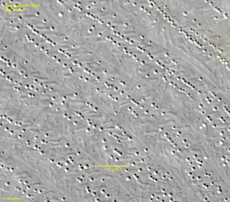
In the pellicle of Spirostomum teres, there are bands of granules (s. fig. 2). Within these bands, the granules partly form orderly rows, which alternate with disorganized areas. However, a rough arrangement of the granules in 3–4 rows can be recognized (s. fig. 3).
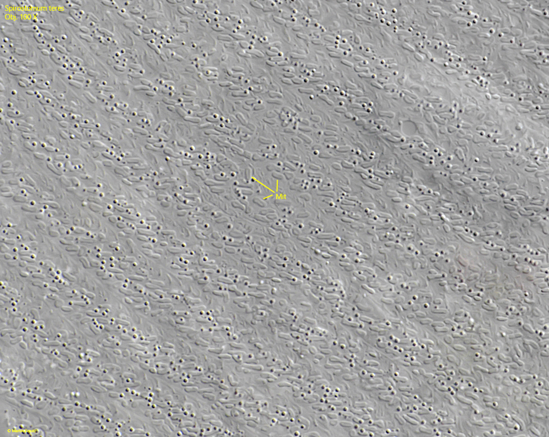
The mitochondria are arranged directly beneath the bands containing granules. They can easily be recognized as short, sausage-shaped structures with a length of 1.5–3.5 µm (s. fig. 4). They provide the energy for the cilia, which are arranged in rows between the bands of granules.
Fig. 1 a-e:Spirostomum teres. L = 650 µm. A freely swimming specimen. Obj. 20 X.
Fig. 2:Spirostomum teres. The rows of sperical granules are arranged between the rows of cilia and runnig spirally counterclockwise to the posterior end. Within the rows the granules are scattered slightly irregularly. Obj. 100 X.
Fig. 3:Spirostomum teres. The cortical granules with a diameter of 0.2–2 µm in detail. Obj. 100 X.
Fig. 4:Spirostomum teres. Beneath the rows of granules the rows of mitochondria (Mit) are visible. They provide the energy for the rows of cilia. The mitochondria have a length of 1.5–3.5 µm. Obj. 100 X.
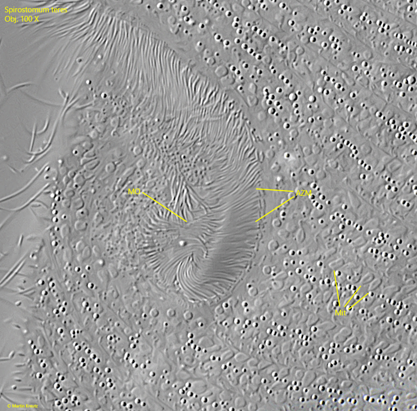
Fig. 5:Spirostomum teres. The adoral zone of membranelles (AZM) on the right side of the oral groove (OG) bend to the right into the cavity of the mouth opening (MO). Obj. 100 X.
Fig. 6:Spirostomum teres. The ellipsoid macronucleus (Ma) with an adjacent, lens-shaped micronucleus (Mi). Obj. 100 X.