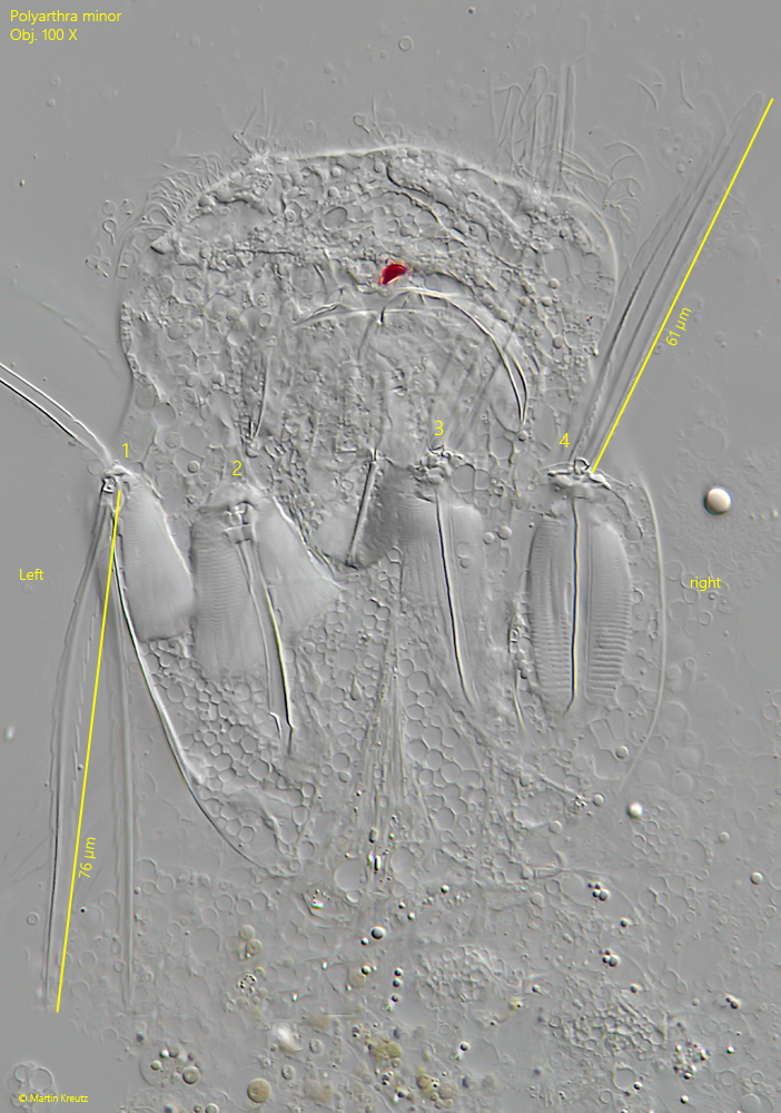
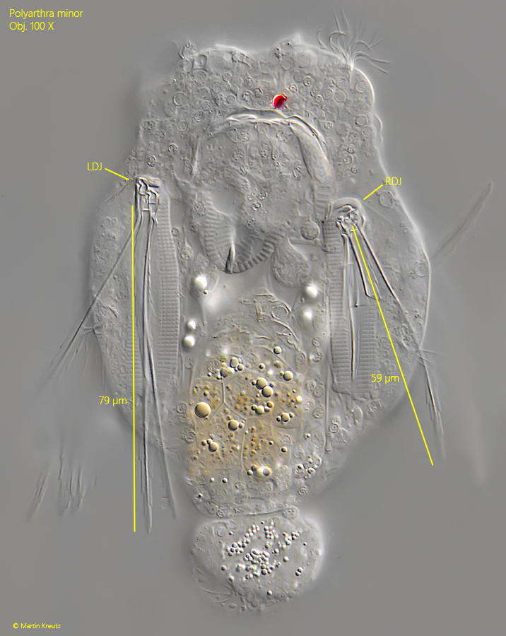
the fins of the left dorsal group significantly longer
dorsal fins very thin
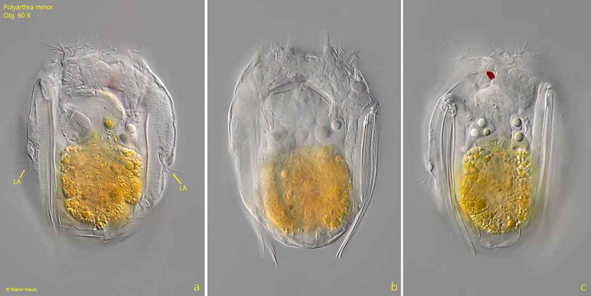
lateral antennae near mid-body
vitellarium with 4 nuclei
one dorsal eyespot
Polyarthra minor
So far I have found Polyarthra minor only in the Sima Moor and in the Diebelsee near Johannisthal. Both bodies of water have a moor-like character with a lot of Sphagnum moss. This is the preferred habitat, as also described in the literature.
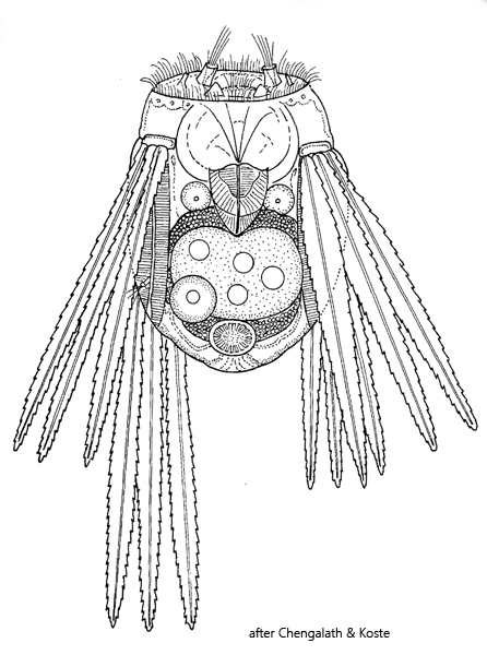
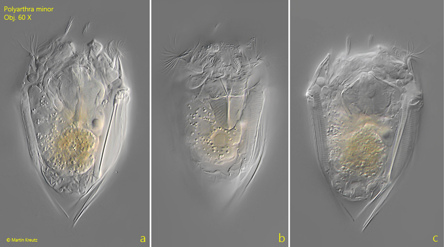
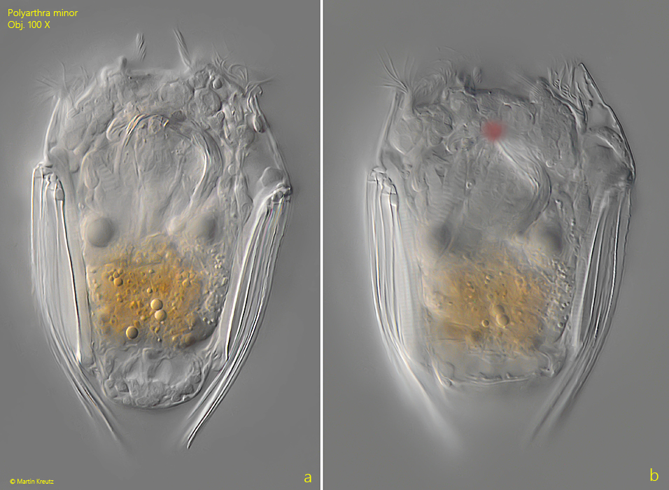
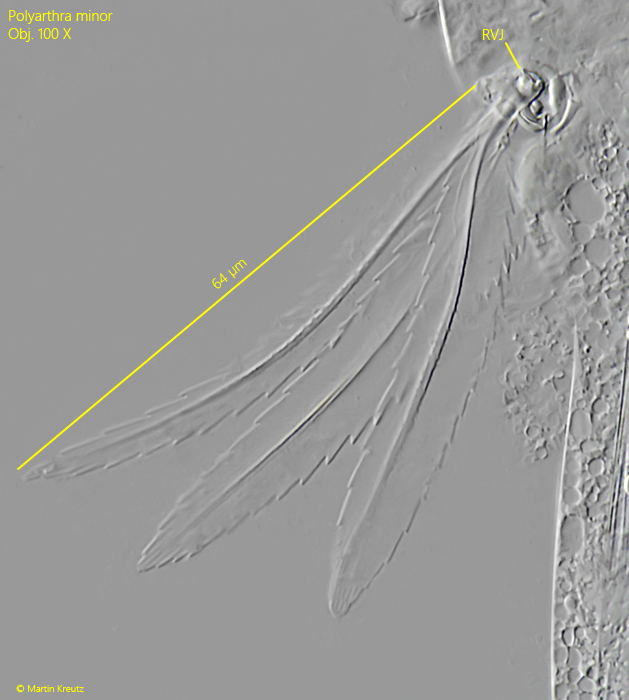
Polyarthra minor is the smallest representative within the genus Polyarthra, with a body length of approx. 80 µm. In the samples, the specimens are easily recognized by the typical body shape. This is approximately rectangular, with the folded lateral fins angled roof-like toward the central body axis at the posterior end. On each side of the body there are two bundles of 3 fins each, with each bundle having a joint to which a strong muscle is attached (s. fig. 4). With the muscles that insert at the joint, the fins can be splayed out abruptly, resulting in a very rapid escape movement.
As early as 1904, Voigt noted that the fins of Polyarthra minor exhibit a peculiar asymmetry. The left, dorsal bundle of fins is significantly longer than the fins of the other bundles. In freely swimming specimens this asymmetry is difficult to discern, but in compressed specimens it becomes apparent (s. figs 4 and 5). According to my measurements, the fins of the left dorsal bundle are about 25–30 % longer than those of the other bundles. The reasons for this asymmetry are not clarified.
Fig. 1 a-c:Polyarthra minor. L = 73 µm (of body). A freely swimming specimen from ventral. Obj. 60 X.
Fig. 2 a-c:Polyarthra minor. L = 87 µm (of body). A second freely swimming specimen from dorsal. Note the lateral antennae (LA) near mid-body. Obj. 60 X.
Fig. 3 a-b:Polyarthra minor. L = 78 µm (of body). A third freely swimming specimen from ventral. Obj. 60 X.
Fig. 4:Polyarthra minor. A strongly squashed specimen from dorsal. The joints (1–4) of the 4 groups of fins are visible with the mucles below. The fins of the left dorsal group (joint 1) are 15 µm longer than the fins on the right side (joint 4), what corresponds to a difference of 25 %. Obj. 100 X.
Fig. 5:Polyarthra minor. A second squashed specimen from dorsal. The fins of the left dorsal joint (LDJ) are 20 µm longer than the fins of the right dorsal joint (RDJ) what corresponds to a difference of 29 %. Obj. 60 X.
Fig. 6:Polyarthra minor. The fins of the ventral bundles are broader compared to the dorsal fins. RVJ = right ventral joint. Obj. 100 X.
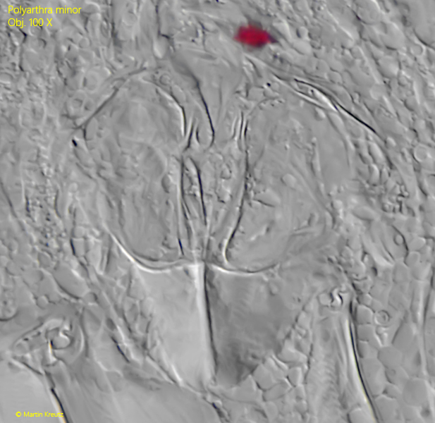
Fig. 7:Polyarthra minor. The trophi in a strongly squashed specimen. Obj. 100 X.