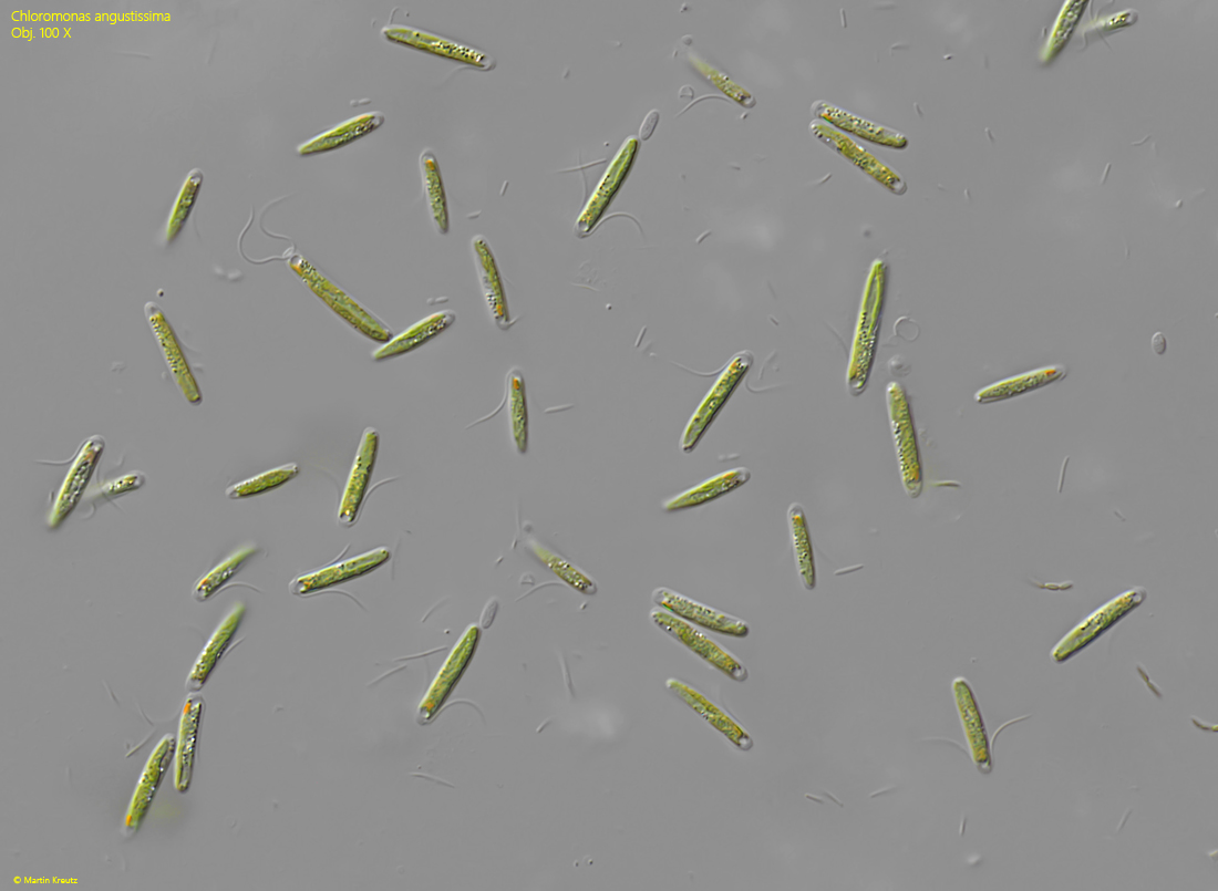
I occasionally find Chloromonas angustissima in the Simmelried. Most of the specimens are located among decomposed plant material. In the fresh samples the specimens are difficult to detect due to their small size. The cells, however, like to settle on the floating coverslip and there they also reproduce to larger populations (s. fig. 1).
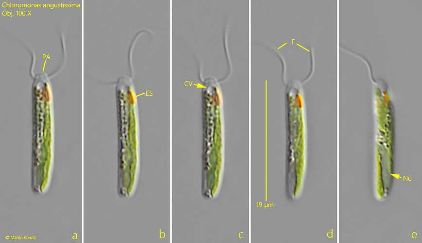
Chloromonas angustissima immediately stands out because of its unusually slender cell shape. At the anterior end there is a transparent papilla and two flagella of equal length (s. figs. 2 a and 2 c). The cells only swim short distances and then rest again, during which the flagella take on a typical S-shape. Only one groove-shaped chloroplast is present. A pyrenoid is absent. Also striking is the orange eyespot at the front end, which is quite large (s. fig. 2 b). I was not able to observe any cell division or the formation of autospores.
Ettl (1958) gave the length of Chloromonas angustissima as a maximum of 8 µm. In my population the cells were 14–21 µm long. All other characteristics, however, correspond to the description of Chloromonas angustissima.
Fig. 1:Chloromonas angustissima. L = 14–21 µm. A collection of specimens on the floating coverslip on a sample from the Simmelried. Obj. 100 X.
Fig. 2 a-e:Chloromonas angustissima. L = 19 µm. Different focal planes of a specimen. Note the papilla (PA) at the anterior end and the small contractile vacuole (CV) below the papilla. ES = eyespot, F = flagella, Nu = nucleus. Obj. 100 X.