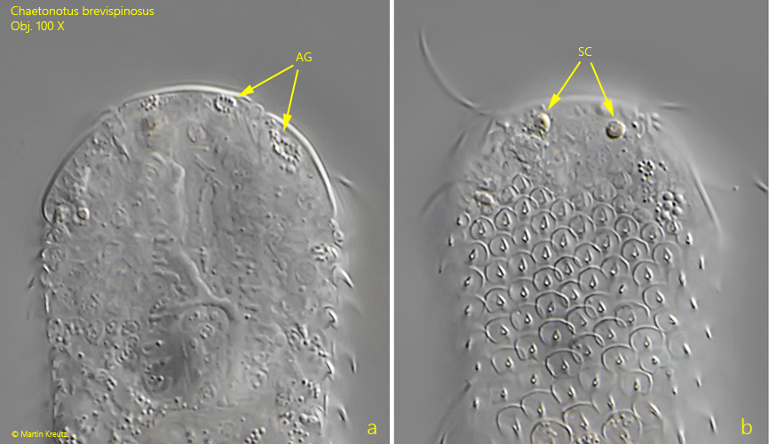
anterior accumulations of granules and “skin cells”
hypostomion absent
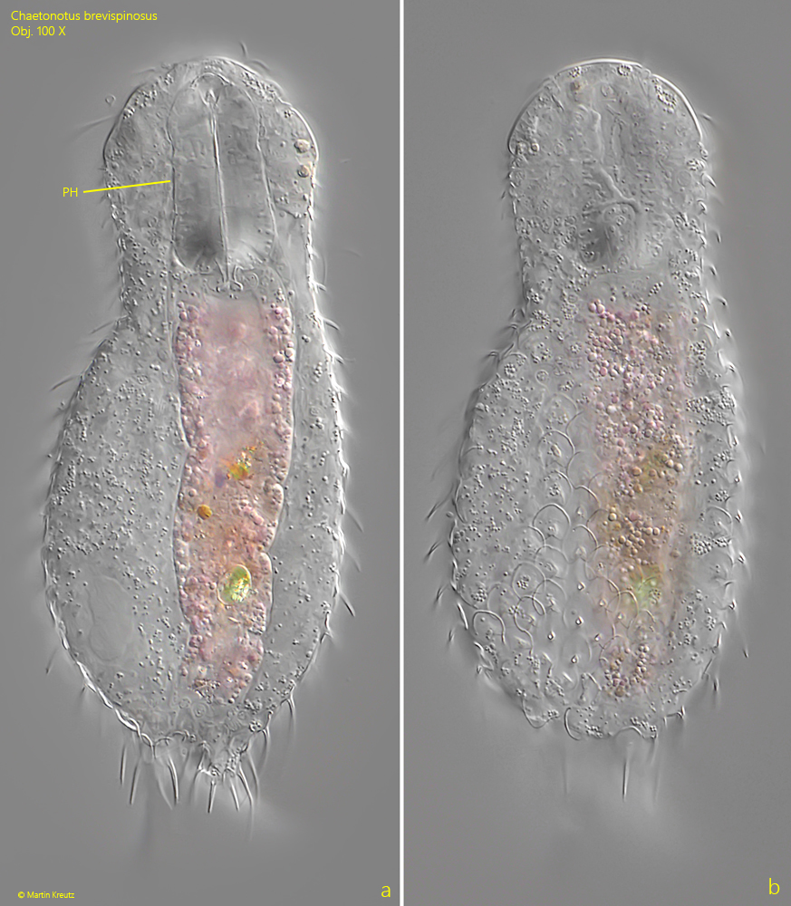
pharynx conical, 20–40 µm long
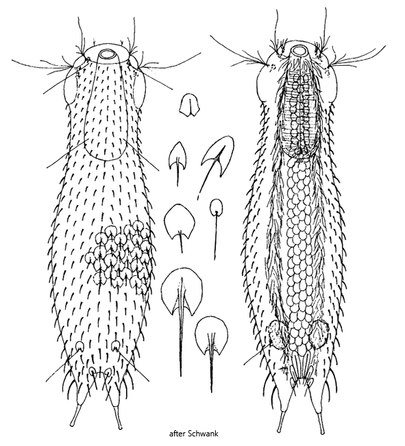
dorsal 11–13 columns of scales
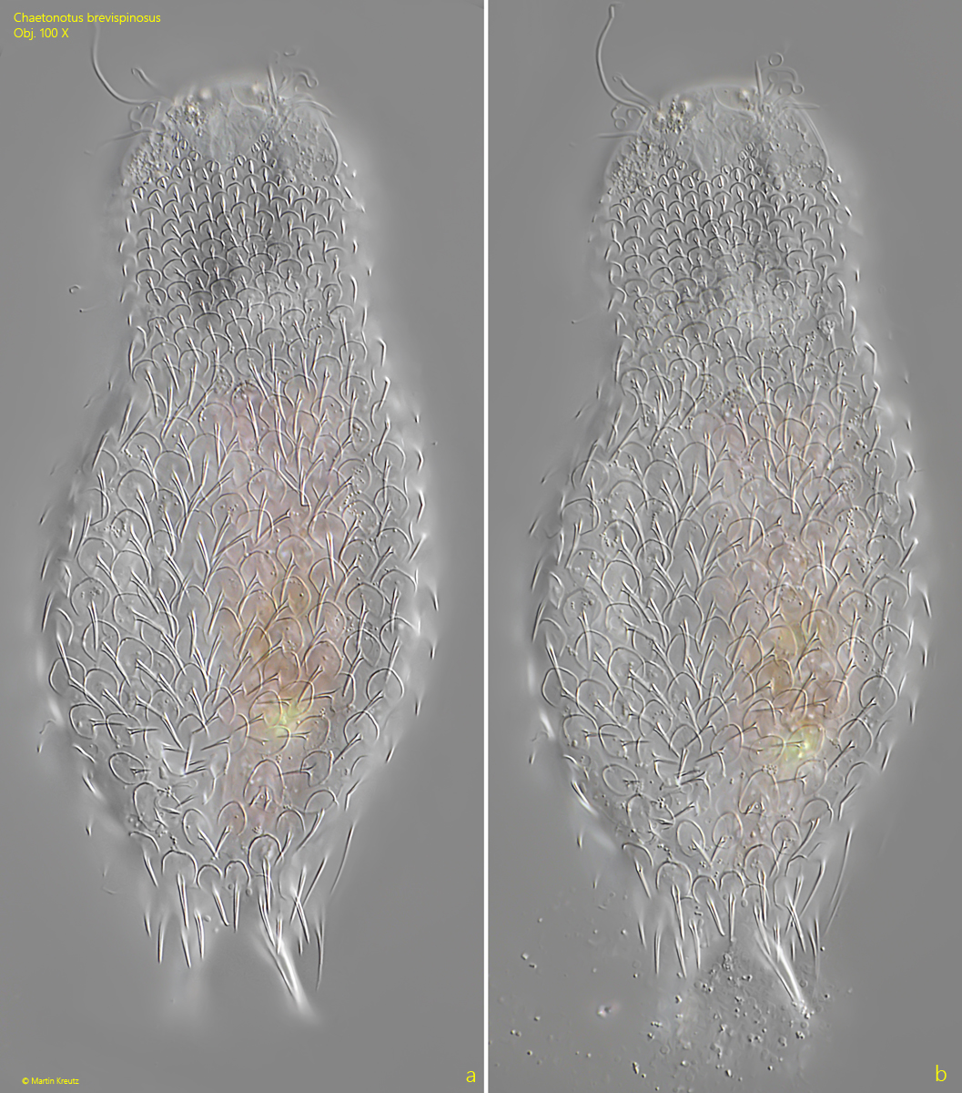
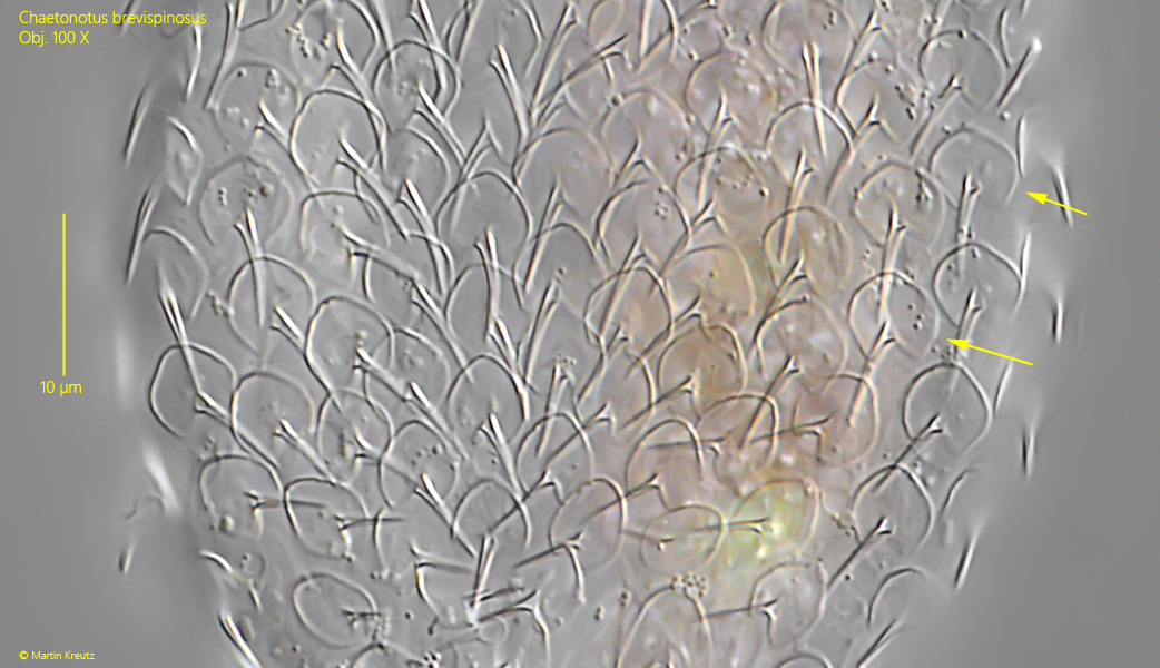
dorsal scales circular, with V-shaped incision, 2–6 µm long
dorsal spines simple, angular, slightly curved
base of toes with 3–5 pairs thickened and elongated spines
ventral 3–5 rows smooth circular scales
two elongated terminal scales and two pairs keeled scales
furca 11–24 µm long, tubes 9–10 µm
Chaetonotus brevispinosus
Chaetonotus brevispinosus is described as a very common and widespread species. So far, however, I have only been able to find a single specimen in July 2023 in the Ulmisried. The specimen was already under the coverslip when I found it. Therefore, the images do not show a free‑swimming specimen.
With a length of about 120 µm, Chaetonotus brevispinosus is a comparatively small species. For identification it is absolutely necessary to analyze the shape of the scales, which can only be seen at high magnification. In Chaetonotus brevispinosus the dorsal scales are almost circular with a length of 2–6 µm. They have a distinct distal notch, which was V‑shaped in my specimen (s. fig. 4). However, this notch can be variable. The scales bear a strong simple spine. The spines are slightly curved and about 5–7 µm long.
Since I found only this one specimen under the coverslip, I was unable to examine the ventral side. According to Schwank (1992) there are several rows of rounded scales, which are sometimes keeled. At the posterior end there are two short-spined terminal scales, which are accompanied by two pairs of keeled scales.
Chaetonotus brevispinosus possesses neither ocelli nor ocellar granules. However, at the anterior margin of the head one can see accumulations of granules and so‑called “skin cells”, which were already described by Remane in 1936. In my specimen I was able to discern both the granules and the “skin cells” (s. fig. 2 a-b). The function of these granules and cells is unknown.
Fig. 1 a-b:Chaetonotus brevispinosus. L = 126 µm. Two focal planes of a slightly squashed specimen. PH = pharynx. Obj. 100 X.
Fig. 2 a-b:Chaetonotus brevispinosus. Two focal planes of the head with accumulations of granules (AG) and so called “skin cells” (SC). Obj. 100 X.
Fig. 3 a-b:Chaetonotus brevispinosus. L = 126 µm. The pattern of the dorsal scales with thick, simple spines. Obj. 100 X.
Fig. 4:Chaetonotus brevispinosus. The dorsal scales in detail. They have a circular shape with an distal V-shaped incision (arrows). Obj. 100 X.