I find Raphidocystis tubifera exclusively in the pond of the campsite Fliesshorn. The species can be found there in the plankton all year round. However, the specimens are never abundant in the samples. The pond is highly eutrophic and must be aerated during the summer months to prevent the formation of anaerobic layers.
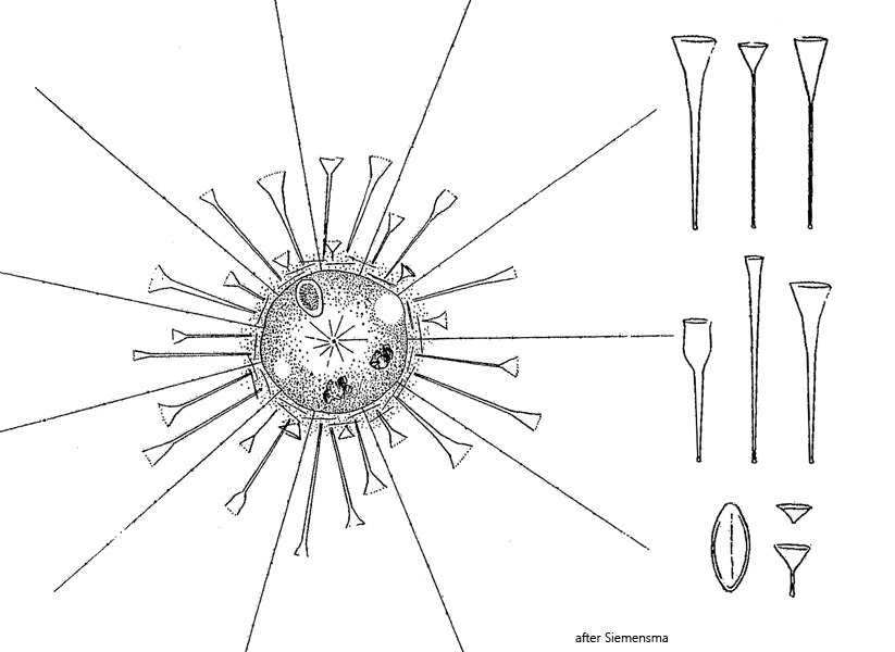
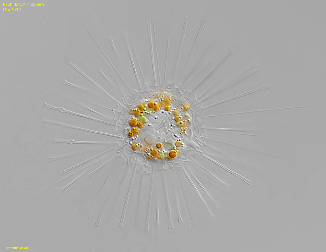
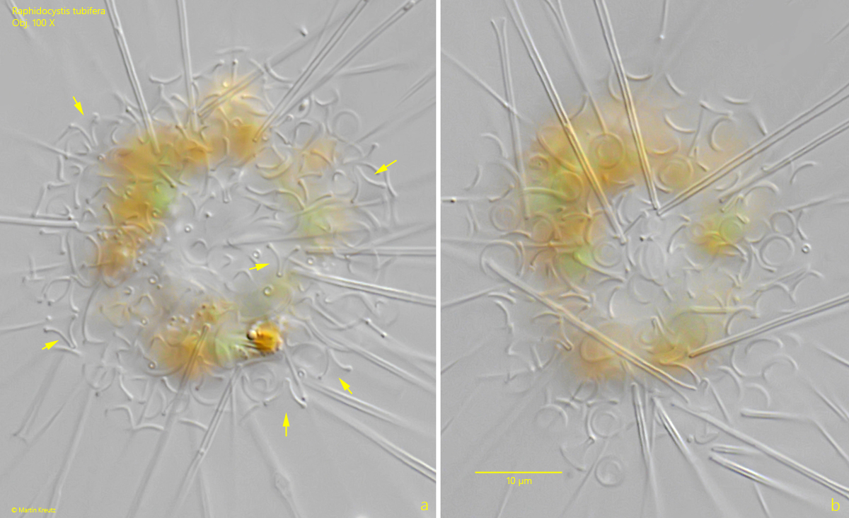
Raphidocystis tubifera cannot be confused with any other species, because of the three different types of scales covering the cell body, the slender, trumpet-shaped scales are very prominent and readily visible even at medium magnification. They project radially from the cell body (s. fig. 1). Directly on the cell body is a layer of oval, tangentially arranged scales, with a layer of funnel-shaped scales above it. The funnel-shaped scales lie irregularly and are not radially aligned. In apical view, their round rim can be seen (s. fig. 3 a-b). The tangential scales have a thickened rim and a central rib, which is difficult to discern (s. fig. 4). Under an electron microscope, both the funnel-shaped scales and the tangential scales show a complex fine structure.
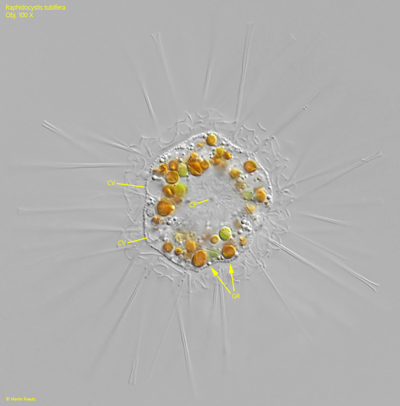
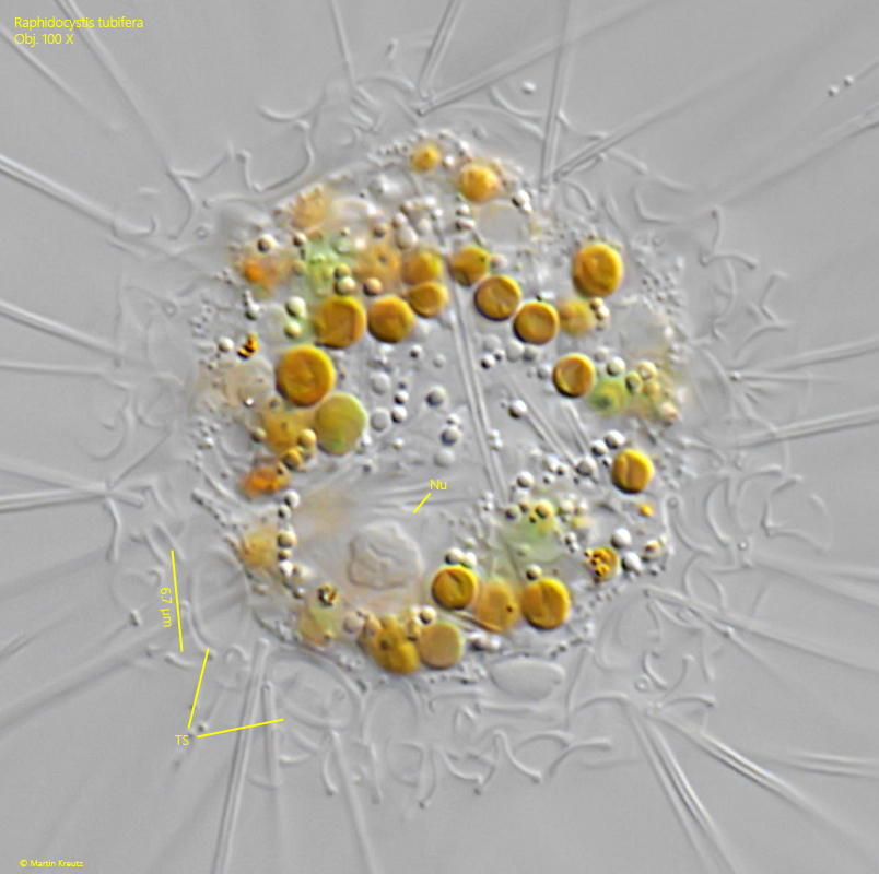
The cell membrane of Raphidocystis tubifera has a layer of fine granules. The cell nucleus is located eccentrically (s. fig. 4), and the centroplast is found in the middle of the cell (s. fig. 2). The centroplast forms and organizes the microtubules with which the axopodia are constructed. Several contractile vacuoles are located at the margin of the body (s. fig. 2).
The cell bodies of the specimens in my population all had a diameter of more than 30 µm and were therefore about 10–20% larger than stated in the literature. The dimensions of the three different types of scales also varied from the values reported in the literature. However, I attribute this to normal variability as well as to the respective habitat.
Fig. 1:Raphidocystis tubifera. D = 35 µm (without coat of scales). A specimen found in October 2026. Obj. 100 X.
Fig. 2:Raphidocystis tubifera. The same specimen as shown in fig. 1 slightly squashed. Two of the contractile vacuoles (CV) are visible and the centroplast (CP). The surface of the cell is covered with fine granules (GR). Obj. 100 X.
Fig. 3 a-b:Raphidocystis tubifera. Two focal planes on the coat of scales. In fig. 1 a several of the funnel-shaped scales are visible in lateral view (arrows). In apical view these scales appear as circles (b). The height of the funnels in this specimen is 3.5–3.8 µm. Obj. 100 X.
Fig. 4:Raphidocystis tubifera. A squashed specimen with focal plane on two tangential scales (TS) with an oval shape. Both scales have a length of 6.7 µm. The eccentrically located Nucleus (Nu) is also visible. Obj. 100 X.
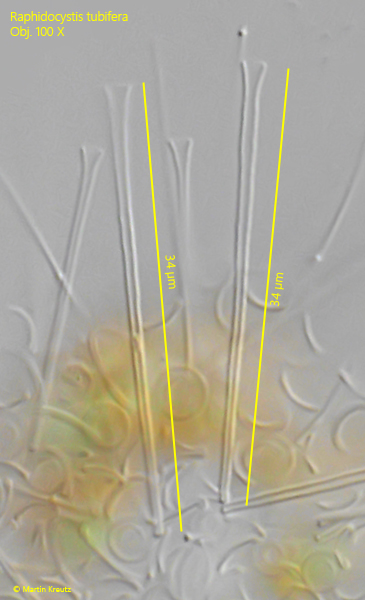
Fig. 5:Raphidocystis tubifera. The slender, trumpet-shaped scales in detail. In this specimen they have a length of 34 µm. Obj. 100 X.