I often find Euglypha tuberculata in the Simmelried. Mostly in the uppermost layer of sediment. Sometimes also in water squeezed from Sphagnum moss.
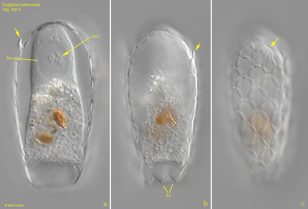
The shell of Euglypha tuberculata is composed of broadly elliptical scales that overlap one another (s. fig. 1 c). They have a diameter of 6–7 µm. They consist of silicate. Triangular, serrated scales surround the circular mouth opening (s. figs. 1 b and 3). In some specimens from my population, the scales at the posterior end had tooth-like extensions (s. fig. 1 a-c). In the literature, I could not find any comparable findings or descriptions of this type of scale in Euglypha tuberculata.
The nucleus of Euglypha tuberculata is usually clearly visible, as it lies in the posterior third of the cell, which usually remains transparent even when there are many food vacuoles in the cytoplasm. In some nuclei of Euglypha tuberculata, I could make out structures that I believe to be condensed chromosomes (s. fig. 1 a). They may have been specimens in which cell division was being prepared.
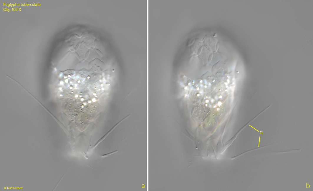
Often there is a layer of highly refractive granules in the middle of the cells (s. fig. 2 a-b). This layer acts as a barrier to food vacuoles, which are mostly located in the anterior part of the cell. The cytoplasm in the anterior half of the cell also often appeared foamy to me, dotted with many small vacuoles. Specimens with extended filopodia are rarely found under the coverslip. According to my observations, they grow to about twice the length of the body (s. fig. 2 a-b).
Fig. 1 a-c:Euglypha tuberculata. L = 60 µm. Three focal planes of a retracted specimen. Note the oval nucleus (Nu) in the anterior third. The structures in the nucleus can be condensed chromosomes (Chr?). The oral aperture is surrounded by serrated scales (AS). Some of the posterior scales show teeth-shaped extensions (arrows). Obj. 100 X.
Fig. 2 a-b:Euglypha tuberculata. L = 57 µm. A specimen with extended filopodia (Fi). Obj. 100 X.
Fig. 3:Euglypha tuberculata. The serrated apertural scales in detail. Obj. 100 X.