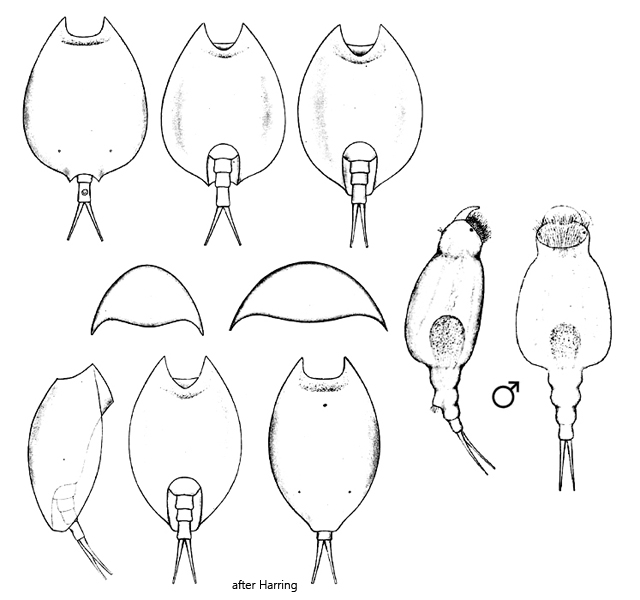
dorsal sinus broadly U-shaped, without granulated fringe
ventral sinus U-shaped without granulated fringe
dorsal end elongated with rounded corners
foot groove U-shaped
foot of three segments, terminal segment longer
toes slender and pointed
Lepadella patella
Lepadella patella is one of the most common rotifers that I find at many of my sampling sites. The specimens are usually found among aquatic plants or on the bottom mud.
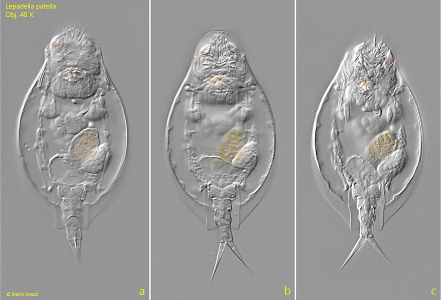
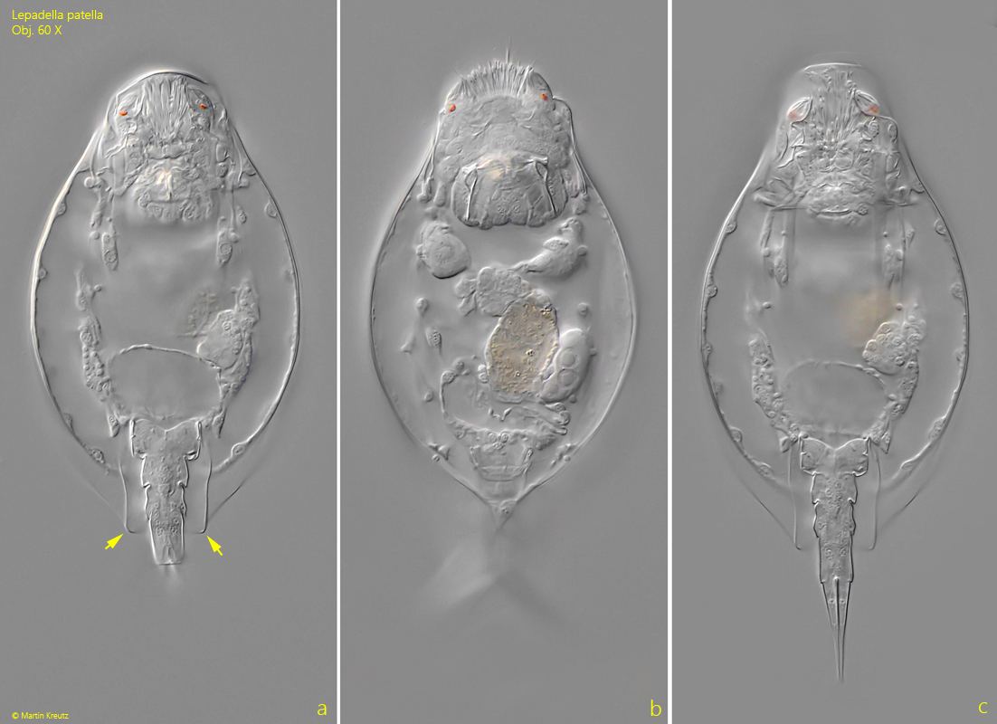
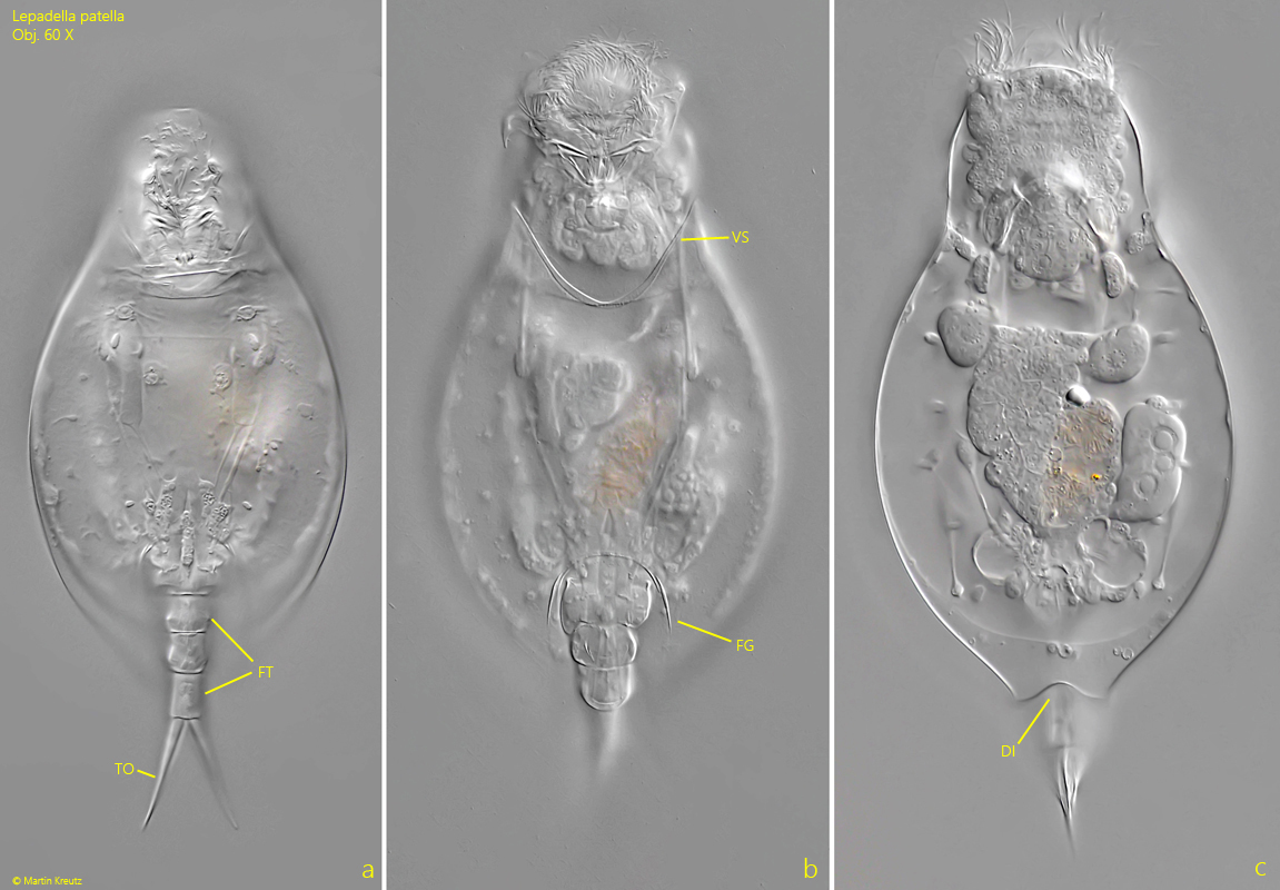
The key characteristics of Lepadella patella are the smooth, convex dorsal side of the lorica, which has no spines or folds. At the posterior end, the lorica is slightly extended dorsally and may have rounded corners, so that it looks almost somewhat angular (s. fig. 2 a). In addition, the posterior margin is often slightly indented (s. fig. 3 c). The ventral side is flat and smooth. The ventral sinus is U-shaped and has no granular collar (s. fig. 3 b), which is an important difference from the similar species Lepadella ovalis. The foot opening is also U-shaped and has no ornamentation (s. fig. 3 b). The foot has three segments, with the distal segment being somewhat elongated (s. fig. 3 a). The toes are slender and taper to a point. They can spread apart and appear pincer-shaped.
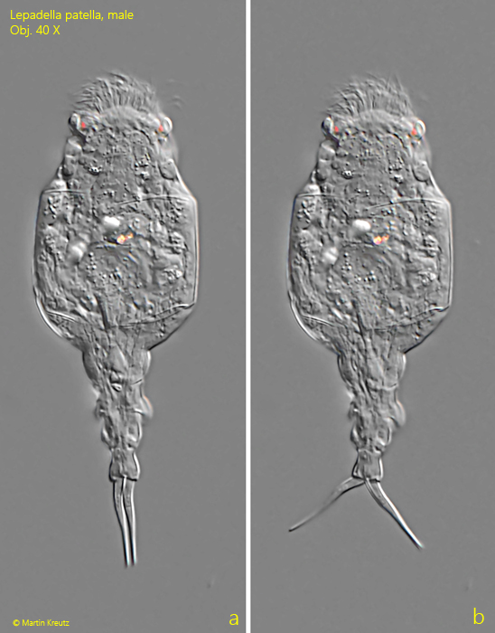
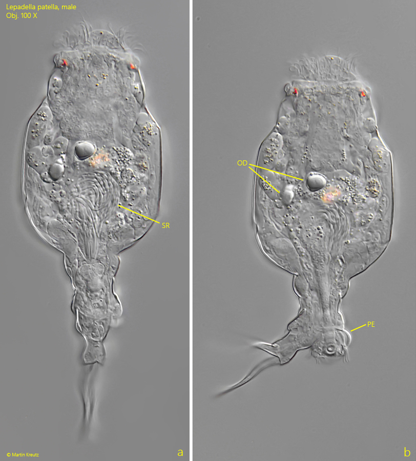
In October 2024, I was able to find a large population of Lepadella patella in a cattle watering trough on the island of Hiddensee. Among the (female) specimens, I was also able to find numerous males, which are quite large at 100–120 µm in total length (s. figs. 5 a-b and 6 a-c). Unlike the female specimens, they have no lorica and no digestive tract. Instead, a large seminal reservoir is found in the middle of the body (s. fig. 6 a). The penis is located on the first segment of the foot (s. fig. 6 b).
Fig. 1 a-c:Lepadella patella. L = 117 µm (of lorica). A freely swimming specimen from ventral. Obj. 40 X.
Fig. 2 a-c:Lepadella patella. L = 117 µm (of lorica). The same specimen as shown in fig. 1 a-c at higher magnification from ventral. Note the rounded corners at the posterior end of the lorica (arrows). Obj. 60 X.
Fig. 3 a-c:Lepadella patella. L = 117 µm (of lorica). A specimen from ventral (a, b) and with focal plane on the dorsal posterior end. The ventral sinus (VS) as well as the foot groove (FG) are U-shaped. The posterior dorsal margin has a slight indentation (DI). FT = foot, TO = toes. Obj. 60 X.
Fig. 4:Lepadella patella. L = 117 µm (of lorica). A squashed young and transparent specimen from ventral. BL = bladder, ES = eyespot, GG = gastric glands, Int = intestinum, MA = mastax, ST = stomach, Vit = vitellarium. Obj. 100 X.
Fig. 5 a-b:Lepadella patella. L = 114 µm (total length). A slightly sqaushed male specimen from ventral. Obj. 40 X.
Fig. 6 a-b:Lepadella patella. L = 114 µm (total length). The same male specimen as shown in fig. 5 a-b at higher magnification. Note the sperm reservoir (SR) in the middle of the body and the penis (PE). OD = oil droplets. Obj. 100 X.
Fig. 7 a-b:Lepadella patella. Two focal planes of the trophi in a strongly squashed specimen. Obj. 60 X.